Journal of
eISSN: 2373-6437


Research Article Volume 5 Issue 3
1Chief Consultant and HOD Institute of Pulmonary Medicine and Research Sri Ramakrishna Hospital, India
2Professor and HOD Department of Cardio-Respiratory Phsyiotherapy College of Physiotherapy, India
3Post graduate Student College of Physiotherapy, India
Correspondence: Mohankumar Thekkinkattil, Chief Consultant and H.O.D Institute of Pulmonary Medicine and Research Sri Ramakrishna Hospital Coimbatore, Tamil Nadu, India
Received: May 27, 2016 | Published: August 5, 2016
Citation: Thekkinkattil M, Muthukumar TS, Monisha R (2016) Influence of Different Body Positioning on Dynamic Lung Functions in Chronic Obstructive Pulmonary Disease Patients and in Normal Subjects-A Comparative Study. J Anesth Crit Care Open Access 5(3): 00189. DOI: 10.15406/jaccoa.2016.05.00189
Background and purpose: Chronic obstructive pulmonary disease represents a substantial economic and social burden throughout the world. Along with pharmacological interventions, all current treatment guidelines emphasize the role of pulmonary rehabilitation in COPD subjects for making them fit at their physical performance and activities of daily living but current treatment guidelines does not emphasize much about the role of body positioning in COPD subjects . The objective of this study was to determine the effect of different body positioning on dynamic lung functions in COPD and in normal subjects.
Methodology: Two groups consisting of 15 COPD and 15 normal subjects aged 40 to 65 years participated in the study. Their dynamic lung functions including FEV1, FEV6, FEV1/FEV6 and PEFR were measured in randomized order in different body positions i.e. standing, sitting, 3/4 sitting, long sitting, supine lying, sidelying right, sidelying left and headdown position.
Results: For all the lung functions, the calculated ‘F’ values when measured showed larger values than tabulated values in COPD and in normal groups.
Conclusion: It is concluded that there is significant difference in the effect of different body positioning on dynamic lung functions in COPD and normal subjects with the maximum results in standing and least in headdown position.
Keywords: Body Positioning; Dynamic lung functions; COPD
Global Initiative for Chronic Obstructive Lung Disease (GOLD) defined COPD as a disease state characterized by airflow limitation that is not fully reversible and is usually progressive, associated with an abnormal inflammatory response of the lungs to noxious particles and gases.1 In the Confronting COPD survey, 80% of patients had two or more symptoms on most or all days, such as breathlessness (45%), cough (46%) and sputum production (40%).2 The World Health Organization estimates that COPD is the fourth leading cause of death worldwide, with 2.74 million deaths in 2000,3 and is projected to rank 5th in 2020 as a worldwide burden of disease.4 The prevalence of physiologically defined chronic obstructive disease in adult aged ≥40 yrs is 9-10%.5
Airflow limitation is the slowing of expiratory airflow as measured by spirometry, with a persistently low forced expiratory volume in the first second (FEV)1 and a low ratio of FEV1–to forced vital capacity (FVC),not reversible with treatment.6 The current GOLD and American Thoracic Society/European Respiratory Society definition of airflow limitation is an FEV1/FVC of < 70% measured with post bronchodilator lung function.1,7,8 Recent well accepted treatment guidelines for COPD urges the use of spirometry and reversibility testing for diagnosis and monitoring.9 Spirometry is a physiological screening test of general respiratory health that measures how an individual inhales or exhales volumes of air as a function of time. The primary signal measure in spirometry may be volume or flow.10 The GOLD definition of COPD classified reversibility as an FEV1 increase of 200mL and 12% improvement above baseline FEV1 after either inhaled corticosteroids or bronchodilators.11
A physiological variable- the FEV1 is often used to grade the severity of COPD.12 FEV1 is central to definition of COPD and classification of its severity. Consequently FEV1 and its change over time are important outcomes in COPD and valuable measures for the assessment of disease progression.13 Body positioning helps in optimizing O2 transport primarily by manipulating the effects of gravity on cardiopulmonary and cardiovascular functions.14 Body position has been shown to affect lung volumes and muscle biomechanics. Higher lung volumes have been linked with better expiratory muscle length- tension relationship and improved expiratory pressures and flow rates. Peak expiratory flow rate (PEFR) has been used as surrogate measure of cough and huff strength.15 The FEV1 and PEFR are well correlated. Both FEV1 and PEFR are most widely used and reproducible measures of forced expiration.16 Body positioning is used during airway clearance treatments to alter lung volumes, reduce dyspnea,18 and maximize ventilation/perfusion matching.19
Till date limited studies have been done to examine the effect of different body positioning on lung volumes and PEFR in the subjects with COPD. So the present study was undertaken to explore the effects of different body positioning on dynamic lung functions in COPD, so that the physiotherapists can recommend on positional changes that may increase the strength of coughing and huffing to enhance the clearance of mucus and which can be used as a part of home management programme to enhance mucus clearance in COPD subjects making them fit at their physical performance and activities of daily living.
Study was carried out in Sri Ramakrishna hospital, Coimbatore. Non-probability randomized sampling method has been used. 30 subjects were included in the study i.e. 15 COPD subjects and 15 normal subjects with age ranging from 40 to 65 years. Their demographic profile and detailed medical record was collected through assessment.
Inclusion
Subjects were selected for the study if they fulfilled the following criteria:
Exclusion
Instruments and tools
Instruments
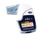
Piko-6 measures FEV1, FEV6 and the ratio of FEV1/FEV6. Piko-6 is very reliable source for measuring lung volumes with a reliability of 0.79.20

The SPIR-O-FLOW measures the peak expiratory flow (PEF) which is the muscular effort to exhale forcibly from fully inflated lungs.
SPIR-O-FLOW meets the new technical standards established by the National Asthma Education Program.

Assessment tool
MRC Dyspnoea Scale
Medical Research Council Dyspnoea Scale has graded the degree of breathlessness related to activities. It has grading from 1 to 5. MRC scale has interrater reliability of 0.92.21
Procedure
Screening of the subjects and allocation of subjects to groups was done by convenient sampling. Informed consent form was signed by the subjects before they participated in the study. Two groups were included in the study: Group A consisted of 15 COPD subjects and Group B consisted of 15 Normal subjects. The subjects were explained what the test will analyze and importance of their involvement for best results. The required procedural details were explained to the subjects and the rest period was given prior to testing.
The subjects were put into the required position, were made comfortable, their clothes were loosened and dentures were removed if they were loose (if present) and were instructed to hold the mouthpiece tightly and seal lips around it and then the subjects breathed into the spirometer (piko-6) and Peak Flow Meter (Spiro-o-flow). In Piko-6, the subjects were instructed to inhale rapidly and completely first and then encouraged to exhale fully and completely and keep breathing out till they can do so no more. Exhalation time suggested by American Thoracic Society (ATS) is 6 seconds unless the subjects cannot or should not continue to exhale further.
The spirometer then digitally displayed the values of FEV1, FEV6 and FEV1/FEV6. The use of nose clip or manual occlusion of nares was done before the subjects started to expire. While in Peak Flow Meter after the point of full lung inflation, subjects were instructed to blow out air vigorously from the mouth without any delay however they need not had to perform the exhalation for 6 seconds. A nose clip is not required for this maneuver. It was checked that there was no false start, no hesitation and cough during the early part of the forced exhalation. It was also checked that adequate inspiration was there before subjects started to expire. Testing was postponed if the subject became short of breath, was too fatigued to continue, could not tolerate the position or was unable to perform the test correctly in that position. In accordance with the recommendations of American Thoracic Society a minimum of 3 trials in each position were obtained for FEV1, FEV6 & PEFR and out of 3 trials the highest value in each position was recorded.
If a variation of 0.150 litres were observed among the largest two values of FEV1 and FEV6, a 4th, 5th and sometimes 6th trial in each position were obtained. And if the largest two out of three acceptable peak expiratory flows were not reproducible with in 40 litres/min upto 2 additional blows were performed.
Each subject was attended for 1 session which lasted for approximately 11/2 hours. Randomization between Piko-6 device and Spir-o-flow and of the different body positions was done by using chit method. Appropriate rest period was given to the subjects after they were put into next position.
The following different positions were used:
Statistical analysis
Data collected was analyzed using ANOVA (software based analysis) to measure the effect of different body positions on dynamic lung function values within the groups (one way ANOVA) and between the groups (two way ANOVA).
Using ANOVA, the Calculated ‘F’ values of FEV1, FEV6, FEV1/FEV6 and PEFR were larger than the tabulated values in COPD and Normal group comparative analysis suggesting that there is significant difference in the effect of different body positions on dynamic lung functions across 2 groups (Figures 1–4).
Evidence show that large number of COPD subjects experience lung volumes and flow rates impairment due to disease process. This abnormal lung function is referred as expiratory airflow limitation. Different body positioning is aimed at maximizing the lung volumes and flow rates which can be utilized to prescribe body positioning in COPD and normal subjects thus increasing expiratory flow rates and volumes which will help in increasing coughing and huffing strength and in turn lead to mucus clearance.
Effect of body positions on these parameters were analyzed using ANOVA. FEV1, FEV6 and PEFR attained the highest values in the standing position followed by sitting having slightly more value than ¾ sitting which in turn is much higher than long sitting followed by supine lying, side lying (Rt.) and side lying (Lt.). Lowest values were observed in head down position. Similar trends were observed in COPD and Normal subjects.
FEV1/FEV6 followed same trend for all the positions in the Normal subjects where as in COPD side lying (Lt.) showed slightly higher values than side lying (Rt.) while all the other positions followed the same fashion. A significant difference in the effect of different body positioning on Dynamic lung functions were observed in COPD and normal subjects. Improvement in dynamic lung functions in more upright positions is noticed in this study which may be due to the reason that in more upright positions gravity pulls the abdominal contents caudally within the abdominal cavity thus increasing the vertical diameter of thorax resulting in increased lung volumes and elastic recoil of lungs, while in recumbent positions the abdominal contents are higher in the abdominal cavity which may interfere with the motion of diaphragm resulting in lower lung volumes and flow rates.
Limitations
Based on the analysis of data it can be interpreted that different body positioning produces significant effect on dynamic lung functions in COPD and normal subjects and can be therapeutically utilized by the COPD and normal subjects to perform expiratory maneuvers to promote secretion clearance and thus improving the general functional well being among themselves.
None.
The authors declare there is no conflict of interests.
None.

©2016 Thekkinkattil, et al. This is an open access article distributed under the terms of the, which permits unrestricted use, distribution, and build upon your work non-commercially.