Journal of
eISSN: 2373-6437


Case Report Volume 9 Issue 1
Professor, Chief of Ear Diseases, Department of the Research Clinical Center of Otorhinolaryngology, Russia
Correspondence: Diab Khassan, Professor, Chief of Ear Diseases, Department of the Research Clinical Center of Otorhinolaryngology, Moscow, Russia, Tel +79191013300
Received: October 29, 2017 | Published: November 14, 2017
Citation: Diab KM, Dayhes NA, Kondratchikov DS, Yusifov KD, Paschinina OA et al. (2017) Cochlear Implantaion in Cases of Inner Ear Pathology. J Anesth Crit Care Open Access 9(1): 00334. DOI: 10.15406/jaccoa.2017.09.00334
The possibility of complications during cochlear implantation (CI) depends on the complexity of the surgical intervention itself, the surgeon’s skill and his experience. The purpose of the study was to improve the surgical stage of CI, taking into account the analysis of the results of reoperations. This study includes 13 revision surgery in pediatric patients after CI with complication. Seven cases of the incomplete electrode insertion into the ossified cochlea and two cases of an electrode extending into the hypotympanum with normal cochlea, were initially identified due to poor auditory skill development or absent behavioral responses following implantation, which prompted imaging. Three cases of the electrode extrusion were initially identified due to otorrhea, patients presented several years after surgery. One case of an electrode placement into the internal auditory canal (IAC) at patient with gusher‒syndrome and Mondini malformation was identified in early postoperative period. Intraoperative findings and management are reviewed. We performed an implant removing and implantation new device (through second turn) at cases of a labyrinthine ossification. Cases of electrode array malpositioning is a rare, but serious and correctable complication in cochlear implant surgery. A multidisciplinary approach, including prompt audiologic evaluation and imaging, is important, particularly when benefit from the implant is limited or absent.
Keywords: Cochlear implantation, Electrode array misplacement, Complications, Ossified cochlea, Inner ear malformation
The inner ear pathology remains one of the most common problem in CI surgery.1,2 Most of the complication after CI surgery main occurs specially in case of inner malformation and ossified cochlea.3,4 The main purpose to determine the features of inner ear structure based on data of preoperatively imaging studies (high‒resolution computer tomography and magnetic resonance imaging) and adequate approach which provide successful surgery and minimize the risk of postoperative complication.5
Describe the cases and causes of incorrect electrode insertion during cochlear implantation and propose an approach to the second cochlea turn in cases of cochlear ossification.
It has been performed 13 revision surgery in pediatric patients after CI between January 2015 and October 2017, at the authors’ institution:3 cases of the electrode extrusion, 7 cases of the incomplete electrode insertion into the cochlea, 2 cases of an electrode extending into the hypotympanum, 1 case of an electrode placement into the internal auditory canal (IAC).
Cases of the extrusion
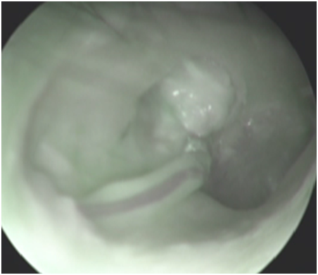
There was electrode array extruding through posterior canal wall 9 years after implantation in first case (Figure 1). It was founded an injury of the posterior wall at the posterior tympanotomy, an epidermis growth over the surface of the electrode in the mastoid cavity during revision surgery. It was performed the electrode extraction, sanitation mastoid cavity, reinsertion of the same electrode and a cartilage electrode covering. There was an electrode extrusion through the tympanic membrane and labyrinthitis in second case. It was founded an injury of the annulus fibrosis. We performed an electrode extraction and tympanoplasty. In 3th case, the extrusion of the electrode array was due to the inflammation in the middle ear caused by Pseudomonas aeruginosa. Explantation and tympanoplasty was performed.

Figure 1 Otoscopic view ‒ electrode array extruding through posterior canal wall 9 years after implantation.
Сases of the incomplete electrode insertion
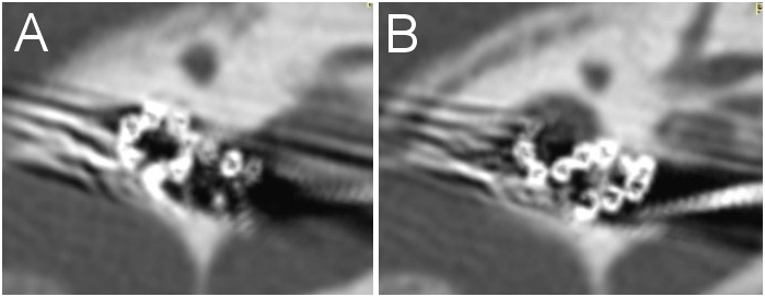
There was 7 cases of a labyrinthine ossification. We performed an implant removing and implantation new device (through second turn) and simultaneously CI at the second ear. The surgical approach was as follows: extended mastoidectomy with exposure of sigmoid sinus, dura mater and sinodural angle. After extended posterior tympanotomy up to the facial nerve canal wall and removal of the incus and bony bridge the first cochleostoma was created in the typical place (with drilling of the basal turn up to its passage into the ascending part taking into consideration risk of internal carotid artery damage). Then the second cochleostomy was performed 1 mm inward to the anterior margin of the oval window niche on the line proceeding from the processus pyramidalis parallel to the stapes crura after removal of the anterior crus of stapes and anterior 2/3 of the footplate. The posterior stapes crus and tendon of stapedial muscle was preserving intact. The insertion of the electrode array was performed through second turn. A remain electrodes placed in exposed basal turn after removal of the part promontory wall separating the basal and the second turn (Figure 2).

Figure 2 CT of left temporal bone. A – Electrode array in second turn. B ‒ Remain electrodes placed in exposed basal turn after removal of the part promontory wall separating the basal and the second turn.
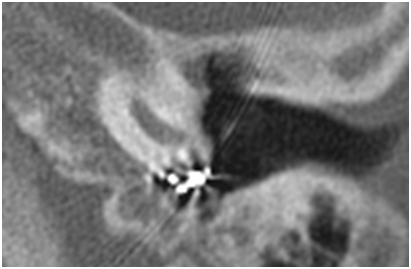
Cases of electrode misplacement into the hypotympanum (Figure 3)
This 2 patents has a rotate cochlea. We extended posterior tympanotomy, extracted an electrode from hypotympanic cells without damage andreinserted correctly the same electrode array via round window.

Figure 3 Axial CT left temporal bone demonstrated the case of insertion of the electrode into the hypothympanum cells.
Case of the electrode placement into the IAC
There was no partition between basal turn of the malformed cochlea (incomplete partition type II) and IAC, electrode was placement through IAC into the cerebellopontine angle (Figure 4). The electrode was being removed, and simultaneous reimplantation of a same implant into the cochlea via an extended same cochleostomy. A postoperative CT scan of temporal bone demonstrated correct electrode placement in the right cochlea (Figure 5).
Based on the study of modern literature, it can be concluded that many authors confine themselves to publishing their own experience in conducting CI. A distinctive feature of our work was the fact that all our patients transferred the primary CI in other clinics to different surgeons, which made it possible to familiarize themselves with the result of the work of several specialists. The reason for the incorrect introduction of the electrode is the presence of one or a combination of several factors. This includes the structure and correlation of the structures of the inner ear, as well as the qualification of the surgeon. In the literature, there are many different options for improper insertion of the electrode into the cochlea. The cases of insertion of the electrode into the upper semicircular canal, into the Eustachian tube, into the hypothympanum cells, into the IAC, into vestibule of the cochlea, displacement of the reference electrode to the dura mater.6 In our work, a rare case of incorrect inserted of an active electrode through the IAC into the cerebellopontine angle in the abnormalities of the inner ear is presented. Extrusion of the electrode into the external auditory meatus is a frequent complication of CI.7 In our opinion, this complication is due to improper administration of posterior tympanotomy, where the posterior wall of the external auditory canal (EAC) is injury. In the process of wound healing and scarring through the created defect of the posterior wall, the electrode prolapses into the EAC. It is also possible to grow epidermis of the skin of the posterior wall of the EAC through a defect along the electrode in the middle ear, with the subsequent formation of cholesteatoma. Given the above, we recommend that at the final stage of the CI, always make an audit of the EAC to ensure the integrity of the posterior wall. Case of exposure of the implant was due to the inflammation in the middle ear caused by Pseudomonas aeruginosa. Some authors assign a key role in exposing the implant body or electrode array to the formation around the last bacterial biofilm, which ultimately leads to explantation of the device.8
The rate of severe complications was low and cochlear implantation is a relatively safe procedure. Routine plain x‒ray an electrode position checks and proper intra‒operative neural response telemetry with good preparation surgeon would avoid rare complications described in this article. Cases that require explantation and reimplantation of the cochlear implant are safe procedures, where the depth of insertion and speech perception results are equal or higher in most cases. Nevertheless, there must be an increasing effort on using minimally traumatic electrode arrays and surgical techniques to improve currently obtained results. Electrode array malpositioning is a rare, but serious and correctable complication in cochlear implant surgery. A multidisciplinary approach, including prompt audiologic evaluation and imaging, is important, particularly when benefit from the implant is limited or absent. Management of electrode arrays in the IAC may be more challenging. Applying the described approach to the second cochlear turn in patients with cochlear ossification enables maximal spiral ganglion cell and modiolus preservation and full electrode insertion, and, therefore, improves postoperative auditory performance.
The Authors report no conflicts of interest. The Authors alone are responsible for the content and writing of the paper.
None.
None.
Authors declare that there is no conflict of interest.

©2017 Diab, et al. This is an open access article distributed under the terms of the, which permits unrestricted use, distribution, and build upon your work non-commercially.