Journal of
eISSN: 2373-6437


Letter to Editor Volume 14 Issue 4
1Department of Anaesthesiology, AIIMS, India
2Department of Anaesthesiology, Saveetha medical college and hospital, India
Correspondence: Dr. Sripriya R, Additional Professor, Department of Anaesthesiology, AIIMS, Mangalagiri, Andhra Pradesh, India, 522501, Tel 9365815939
Received: July 21, 2022 | Published: August 16, 2022
Citation: Dr. Sripriya R, Dr. Surya R. An illustration of the evolution of various injection points for circumventing supraclavicular brachial plexus block failure.J Anesth Crit Care Open Access. 2022;14(4):144‒146. DOI: 10.15406/jaccoa.2022.14.00525
The point of local anesthetic (LA) deposition within the Supraclavicular Brachial Plexus (SCBP) using ultrasound guidance has progressed from “Centre cluster”, “Corner Pocket”, “Double injection” to the “Targeted Intracluster” injection technique to provide a consistently successful block. We have attempted to illustrate the pattern of LA spread with the different injection points using the high-resolution images obtained with the linear‑array transducer (HFL50, 15–6 MHz) of the X‑Porte Ultrasound system (FUJIFILM Sono Site, Inc., Bothell, USA). 12 ml of LA was used for each technique. Prior written informed consent was obtained from the patients for using their sonograms for publication.
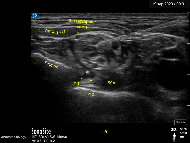
In Figure 1a, the needle tip is positioned in the centre of the “Bunch of grapes” and illustrates the LA spread obtained with the “Centre-cluster” injection technique.1 An important drawback with centre-cluster injection was ulnar sparing. In this image we can well appreciate that though the LA spread has occurred across a major part of the plexus, the two hypoechoic structures representing the neural elements of C8 and T1 roots at the 6’0 clock position to the plexus are spared.

Figure 1a Image depicts the needle tip at the center of the cluster – the ‘Centre-cluster injection’ technique. The local anaesthetic is seen to bathe a major portion of the plexus, but the C8, T1 roots are spared.
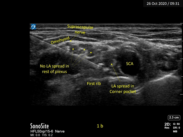
To overcome ulnar sparing, it was suggested to target the “Corner Pocket”.2 Figure 1b illustrates the spread of LA when the needle tip is positioned at the corner pocket. Again, we can observe that the LA has spread across the lower trunk while the rest of the brachial plexus is spared, which explains why a 100% success rate could not be achieved even with this technique.

Figure 1b Image depicts the needle tip at the corner pocket – the ‘Corner- pocket injection’ technique. The local anaesthetic is seen to have spread across the lower trunk while the rest of the plexus looks spared.
The “Double Injection” or “Cluster-Corner” concept then came into vogue, where the LA was injected at both sites thereby taking advantage of both the injection techniques.3
Subsequently, the “Targeted Intracluster” approach was put forth by Techasuk et al. as providing a 100% success rate with a quick onset.4 The availability of high-resolution US imaging was an important contributory factor in delineating several clusters within the “Bunch of grapes” by Techasuk et al. Figure 1c illustrates the uniform distribution of LA across the entire plexus obtained with small aliquots injected into the various clusters. It is however prudent not to miss out on any clusters. At the supraclavicular area, anterior and posterior divisions of the upper trunk may have split away from the main cluster and the suprascapular nerve has also diverged from the rest of the plexus and sparing can occur if any of these clusters are missed.
None.
None.

©2022 Dr., et al. This is an open access article distributed under the terms of the, which permits unrestricted use, distribution, and build upon your work non-commercially.