Journal of
eISSN: 2572-8466


Research Article Volume 5 Issue 5
Department of Pharmaceutical Biotechnology, Malla Reddy College of Pharmacy, India
Correspondence: Rahamat Unissa, Department of Pharmaceutical Biotechnology, Faculty of Pharmacy, Malla Reddy College of Pharmacy, India
Received: August 30, 2018 | Published: October 19, 2018
Citation: Sunil G, Devi PR, Sri GD, et al. Screening of larvicidal activity of nanoparticles synthesized from flower extracts of Hibiscus vitifolius . J Appl Biotechnol Bioeng. 2018;5(5):316-319. DOI: 10.15406/jabb.2018.05.00157
The objective of the present study was to evaluate the larvicidal activity of silver nanoparticles synthesized from aqueous extracts of Hibiscus vitifoliusLinn flowers against the larvae of Aedes aegypti. Stable silver nanoparticles were synthesized by biological reduction method. The parasite larvae were exposed to varying concentrations of aqueous extract of Hibiscus vitifolius flowers and synthesized silver nanoparticles for 24 h as per World Health Organization protocols. Distilled water served as control. Percentage mortality was recorded. The synthesized nanoparticles exhibited significant larvicidal activity. This method is considered as an innovative alternative approach using green nanochemistry technique to control vector parasites and is the first report on mosquito larvicidal activity of Hibiscus vitifolius Linn flowers mediated synthesized silver nanoparticles.
Keywords: Hibiscus vitifoliusLinn, Aedes aegypti, larvicidal activity, silver nanoparticles
Dengue fever is mosquito- borne tropical disease, more common to tropical countries such as India due to the favorable ecological conditions.1 The mosquito Aedes aegypti is a vector for many viral pathogens that causes serious threat to the human beings.2 It is responsible for the transmission of several diseases such as yellow fever, chikungunya and dengue fever etc.2
As there is no specific antiviral drug for the treatment of these diseases, vector control is the best measure.3 To solve this problem, many chemical insecticides may be used to control the mosquitoes, but most of them currently in use are non-selective, poisonous to human health and could also produce environmental contamination.4 Hence there is a need for developing an eco-friendly target specific newer mosquitocidal agents from the natural sources such as plants. Plants constitute a number of chemical substances with medicinal as well as insecticidal activities. More than 2000 plants belonging to different families were found to have potential insecticidal activities. Hibiscus vitifolious is one such plant with unique medicinal activities. In our previous studies, we have reported antimicrobial as well as cytotoxic activities of the plant.5 Here in our present study, we have carried out production of silver nanoparticles (AgNPs) using plant extracts as reducing, stabilizing, and capping agents. This technology involves the usage of microbicidal properties of silver, the insecticidal activity of the selected plant.
Collection of flowers
Fresh flowers of Hibiscus vitifolius were collected from Bahadurpally village, R.R. District, Telangana, India, during the month of October 2017 and identified by Dr. Nirmala Head of the Department, Osmania University, Telangana, India. A Voucher specimen (PH- 806) was deposited in the herbarium of the college.
Rearing of larvae
Water samples were collected from the unused wells near Osmania University, Secunderabad. Huge numbers of larvae and eggs were available in the unused wells, which made it possible for the entire larvicidal assay. Fresh larval forms were reared in the laboratory. Preliminary identifications of the eggs (present in the water samples) were done in Zonal Entomological Research Centre, Secunderabad. In the process of rearing the larval forms of the mosquitoes, the eggs were initially immersed in the 0.01% formaldehyde solution for 30–40 minutes6 (to prevent microsporidian infections), followed by deionized water to facilitate hatching.
After hatching, first instar larvae were distributed in the white plastic cups. Care was taken to prevent overcrowding until development to early 4th instar larvae required for the study. The bacterial broth that consisted of 0.05g of yeast, 0.25g CM0001 nutrient broth in 0.71ml of deionized water was used as natural medium for mass cultivation of the larvae7. Water in rearing container was refreshed every day by removing a little quantity of water from the rearing cups and replacing with fresh water. Different stages of the larval rearing are shown in Figure 1. Further, the larvae were identified based on the macroscopic and microscopic morphological studies (Figure 2), based on the “Identification of the U.S mosquito larvae -manual”.
Preparation of the extract
Flowers of Hibiscus vitifolius were washed thoroughly with autoclaved distilled water and dried in shade for a week and ground using a mixer to the coarse powder. The powder was used for preparing the aqueous extract. 1 g of flower powder was boiled in 10 ml of deionized water for 10 minutes. It was cooled and filtered through Whatman No. 1 filter paper, and the filtrate was stored at 4°C until further use.
Synthesis of SNPs
Silver nitrate (AgNO3) of analytical grade (AR) was purchased from Merck (India). To synthesize silver nanoparticles, 1ml of the aqueous extract of Hibiscus vitifolius flower was added to 100ml of 1mM AgNO3 solution in 150ml glass beaker. Then the beaker was incubated for 24 hrs at room temperature on a magnetic stirrer in the dark place for the reduction of SNPs. The color change from light yellow to dark orange indicated the formation of SNPs. An initial setup was also maintained as flower extract without the addition of AgNO3.
Mosquito larvicidal bioassay
Toxicity of the biologically synthesized nanoparticles towards the larval forms of the mosquito was tested by the standard protocol given by WHO.8 Around twenty (20),4th instar larvae were picked up randomly and were placed into 200ml of sterilized double-distilled water and incubated at 27°C with a photoperiod of 16:8-h light/dark cycle. The effectiveness of silver nanoparticles as mosquito larvicides was determined from all the twenty 4th instar larvae with exposure to time periods.
The larvae were separated into four small specimen bottles containing 25ml distilled water and the larvae were then exposed to each of the concentrations of the extracts in a final volume of 245ml distilled water taken in 500ml bowls (10, 20, 30, 40 and 50mg/l respectively). The nanoparticle solutions were diluted using double distilled water as a solvent according to the desired concentrations (5.0, 4.0, 2.0, 1.0, and 0.5mg/L). The effectiveness of silver nanoparticles as mosquito larvicides was determined from all the twenty 4th instar larvae with exposure to time periods. At each tested concentration, four trials were made and each trial consists of four replicates and the control were tested for anti–larval effects. The larval mortalities were assessed to determine the acute toxicities on 4th instar larvae of Aedes aegypti at intervals of 1, 3, 6, 12,16, and 24 hours of exposure. A number of dead larvae were counted from the 1st hour of exposure, and the percentage of mortality was reported from the average of four replicates. The larval mortality data were corrected for control mortality by the formula of Abbott.9
Since silver nanoparticles are considered as potential targets for various biological applications including antimicrobial, their application as a mosquito larvicidal agent was also investigated. Preliminary phytochemical tests performed on the crude aqueous extract (using standard protocols) showed the presence of proteins, amino acids, steroids, tannins and glycosides.10–12 The results of the screening tests are presented in Table 1.
S. No |
Name of the test |
Aqueous extract |
1 |
Test for Carbohydrates |
_ |
2 |
Test for Proteins and amino acids |
+ |
3 |
Test for Triterpinoids and steroids |
++ |
4 |
Test for Alkaloids |
_ |
5 |
Test for Cardiac glycosides |
+ |
6 |
Test for Anthraquinone glycosides |
+ |
7 |
Test for Saponin glycosides |
+ |
8 |
Test for Flavonoids |
_ |
9 |
Test for tannins and phenolic compounds |
+ |
10 |
Test for fixed oil and fats |
+ |
Table 1 Phytochemical screening of aqueous flower extract of H vitifolia
In the present research green synthesis of AgNPs were carried out using Hibiscus vitifolius flower extract. The change in the color of the aqueous extract to yellowish brown and then to dark brown upon addition of silver nitrate (1m M) solution confirmed the formation of silver nanoparticles. It was noticed that silver ions, when treated with aqueous extract were reduced in solution there by leading to the formation of silver hydrosol. The color intensity was increased with the increase in the incubation time. The time required for the color change differs from to one plant to other. In the present study, Hibiscus vitifolius synthesized SNPs after 24 hrs of reaction. High resolution scanning electron microscopic analysis provided information on the morphology and size of the nanoparticles which was found to be on an average of 35nm.
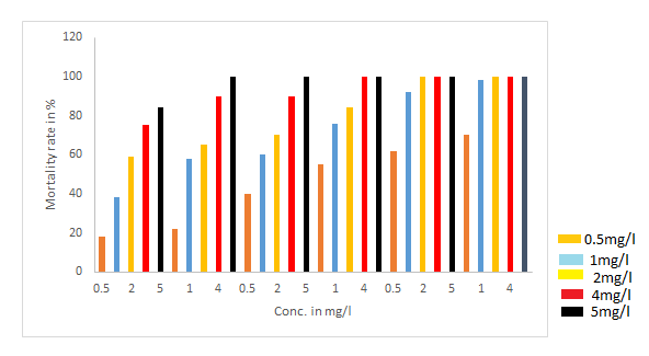
The larvicidal activity of aqueous flower extracts of Hibiscus vitifolius and silver nanoparticles synthesized using Hibiscus vitifolius flowers were tested against 4th instar larvae of the dengue vector Aedes aegypti. The 4th instar larvae thus isolated were identified based on the morphological characteristics and treated with different concentration of aqueous flower extract (10, 20, 30, 40 and 50mg/l) and green synthesized silver nanoparticles (0.5,1,2,4 and 5mg/l). Larvicidal activity was seen in the form of morphological abnormalities. It was observed that larvae treated with high dose levels were reduced in body size and showed incomplete metamorphosis. Stiffness in the cuticle was also observed in a few cases.
The results of the activity are presented in the Figure 3−5. The data obtained from the present study clearly indicate that silver nanoparticles could provide excellent larval control of Aedes aegypti. Greater mortality is seen in larvae treated with silver nanoparticles compared to aqueous extract. It was observed that, aqueous extract and distilled water had little or no effect on larval mortality. Higher concentrations of aqueous plant extract caused no death of the larvae until 12 hours of exposure. The nanoparticle at 1.0 mg/l slightly decreased the survival of larvae to 50% after 12 hours of exposure, while 100% mortality of the larval population was observed in a concentration of 5.0 mg/l nanoparticles within three hours. The nanoparticle of 1.0 mg/l killed the larvae slowly and nearly 90% mortality was found after 16 hours of exposure. The maximum efficacy was observed in 5.0 mg/l of silver nanoparticles.

Figure 3 Survival percentage of mosquito larvae after exposure to different concentrations of silver nanoparticles
The mechanism which causes the death of the larvae could be the ability of the nanoparticles to penetrate through the larval membrane. The silver nanoparticles in the intracellular space can bind to sulphur-containing proteins or to phosphorus containing compounds like DNA, leading to the denaturation of some organelles and enzymes.13,14 Subsequently, the decrease in membrane permeability and disturbance in proton motive force causes loss of cellular function and finally cell death.
The nanoparticles synthesized from the fresh flowers of Hibiscus vitifolius extract showed excellent larvicidal activity against Aedes aegypti. Green synthesis of nanoparticles and its effective use for controlling mosquitoes could help in the development of more potent and environmentally safe mosquitocidal agents. Further studies must be carried out for the isolation and identification of the bioactive compounds and its safety levels.
The authors hereby declare that the work presented in this article is original and that any liability for claims relating to the content of this article will be borne by them.
The authors wish to thank and appreciate the management of MallaReddy College of Pharmacy College for providing necessary facilities to carry out the research work.
The authors declare no conflict of interest.

©2018 Sunil, et al. This is an open access article distributed under the terms of the, which permits unrestricted use, distribution, and build upon your work non-commercially.