Journal of
eISSN: 2572-8466


Research Article Volume 6 Issue 2
1Department of Anatomy, University of Port Harcourt, Nigeria
2Department of Anatomy, University of Ilorin, Nigeria
3Department of Anatomy, Madonna University, Nigeria
Correspondence: John Nwolim Paul, Department of Anatomy, Faculty of Basic Medical Sciences, College of Health Sciences, University of Port Harcourt, Choba, Port Harcourt, Rivers State, Nigeria
Received: January 24, 2019 | Published: March 25, 2019
Citation: Oladipo GS, Paul JN, Amasiatu VC, et al. An examination of carrying angle of students in Madonna University, Elele, Port Harcourt, Rivers State, Nigeria. J Appl Biotechnol Bioeng. 2019;6(2):95-99. DOI: 10.15406/jabb.2019.06.00179
Background: The carrying angle is defined as the acute angle made by the median axis of arm and median axis of forearm in full extension and supination. This angle permits the forearms to clear the hips in swinging movements during walking and is important when carrying is a small degree of Cubitus valgus, formed between the axis of a radially deviated forearm and the axis of the humerus. This study was aimed at examining the carrying angle of male and female students of Madonna University Nigeria.
Materials and methods: The study comprised a total number of 200 subjects (100 male, 100 female), ages between 16 -25years. The carrying angle was measured using digital vernier caliper and compass while height, hip and waist circumference were measured using measuring tape.
Results: Age=21±2.59 years (Male), 20.37±3.00 years (Female); Hip Circumference=90.95±6.63cm (Male), 90.67±8.65cm (Female); Right Carrying Angle=9.31±1.67° (Male), 9.75±2.26° (Female); Left Carrying Angle=8.99±1.53° (Male), 9.58±2.10° (Female); Height=181.09±4.76cm (Male), 175.08±8.34; Waist Circumference=79.72±6.53cm (Male), 82.13±5.85cm (Female). In male subjects, Right Carrying angle can be predicted from other measured parameters as follows [RHLH; RCA=0.1728 (H)-21.991] with a prediction accuracy of 24%; Left Carrying Angle [RHLH; LCA=0.1023 (H)-9.5289] with a prediction accuracy of 10% while in females, Right Carrying Angle can be predicted from other parameters as follows [H; RCA=0.116 (H)-10.559] with a prediction accuracy of 18%; Left Carrying Angle [H LCA=0.1102 (H)-9.7203] with a prediction accuracy of 19%. Independent sample T-test was used to compare differences (sexual dimorphism) in the measured parameters indicated there were significant differences (p<0.05) in all the measured parameters.
Conclusion: The study of carrying angle in Madonna students have shown that the males have mean values (right 9.31 and left 8.99) while the females (right 9.75 and left 9.58). This has also proved that carrying angle is a useful tool in investigating gender variation in forensics and anthropological studies.
Keywords: carrying angle, hip circumference, waist circumference, height, age
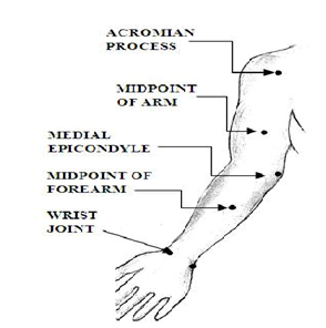
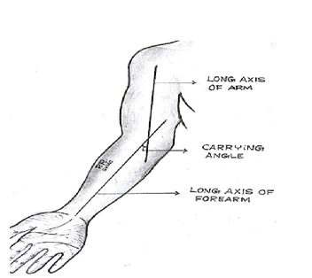
The carrying angle is defined as the acute angle made by the median axis of arm and median axis of forearm in full extension and supination. This angle permits the forearms to clear the hips in swinging movements during walking and is important when carrying is a small degree of Cubitus valgus, formed between the axis of a radially deviated forearm and the axis of the humerus. It helps the arms to swing without hitting the hips while walking. Normally it is 5-15o away from the body or 165-175o towards the body. A decreased carrying angle can result in the forearm pointing towards the body, known as gunstock deformity or cubitus varus.1–7 In Figures 1 & 2 below, the landmarks in the upper limbs and the axes of carrying angle are shown.

Figure 1 Indicating the landmarks in the upper limbs.5

Figure 2 Indicating the landmarks in the the axes of carrying angle.5
Brief anatomy of the elbow joint
The elbow is a complex synovial joint formed by the articulations of the humerus, the radius and the ulna. The elbow joint is made up of three articulations
Movements
The elbow is a trochoginglymoid (combination hinge and pivot) joint.8
Ligaments
Joint capsule
The study was descriptive and comprised a total number of 200 subjects (100 male, 100 female), ages between 16 -25years. All subjects used for this study were from Madonna University, Elele Rivers State, Nigeria. They were healthy individuals free of congenital or acquired abnormalities, and trauma. Ethical clearance was obtained from the Research Ethics Committee of the Madonna University, Elele, Port Harcourt, Rivers State, Nigeria.
A thorough clinical examination of the elbow region was done by following all the inclusion and exclusion criteria. The carrying angle (CA) was measured using clinical method on both upper limbs. Points were made 5cm above and below in line with the medial epicondyle in the front of arm and forearm. Width of the arm, forearm and wrist were measured with the help of the Digital Vernier Caliper, with the prongs of the caliper just touching the skin without giving any pressure to derive their midpoints. Two axes were drawn, one from acromian process meeting the midpoint in front of arm, another in forearm joining the midpoints in front of forearm and wrist. Both lines were extended so that they intersect nearly in front of the elbow joint and the angle thus formed in the medial aspect represents was measured using a compass.
Distribution of carrying angle and other parameters was done using descriptive statistics, Independent sample T-test guided by Levene’s test for Equality of Variance was used to evaluate sex based differences, while Paired sample T-test was used to determine side differences (left and right). Correlation and regression analysis was done, generating a regression equation for estimating the Height of subjects from their Hand parameters. Significance level was set at 95% confidence interval, hence P< 0.05 was considered significant. All these were carried out with the aid of the Statistical Package for the Social Sciences (SPSS IBM® ver 23.0) and MS Excel.
In Table 1, the descriptive statistics of hand dimensions were as follows (Age=212.59 years (Male), 20.373.00 years (Female); Hip Circumference=90.956.63cm (Male), 90.678.65cm (Female); Right Carrying Angle=9.311.67 (Male), 9.752.26 (Female); Left Carrying Angle=8.991.53 (Male), 9.582.10 (Female). In Table 2, Independent sample T-test was used to compared significant differences (sexual dimorphism) in the measured parameters. Significant differences were found in all the measured parameters. In Table 3, Side differences (left and right) were determined using paired sample T-test. Significant difference was found in male subjects between the right and left carrying angle (t=2.62, P=0.01), but not in female subjects (t=2.02, P=0.05) at P< 0.05. In Table 4, there was a strong correlation between carrying angles in males and females with Hip circumference, Waist circumference, Height. In Table 5, a summary of correlation and regression analysis was presented, with a regression equation for estimating the carrying angle of subjects from other parameters (Height, Hip Circumference, Waist Circumference and Age).
Parameters |
MALE (N = 100) |
FEMALE (N = 100) |
TOTAL (N = 200) |
|||||||||
Min |
Max |
Mean |
S.D |
Min |
Max |
Mean |
S.D |
Min |
Max |
Mean |
S.D |
|
Age(years) |
17.00 |
28.00 |
21.33 |
2.59 |
17.00 |
29.00 |
20.37 |
3.00 |
17.00 |
29.00 |
20.85 |
2.84 |
Hip Circumference (cm) |
75.00 |
112.00 |
90.95 |
6.63 |
73.50 |
115.00 |
90.67 |
8.65 |
73.50 |
115.00 |
90.81 |
7.69 |
Right Carrying Angle (°) |
5.00 |
12.00 |
9.31 |
1.67 |
5.00 |
17.00 |
9.75 |
2.26 |
5.00 |
17.00 |
9.53 |
1.99 |
Left Carrying Angle (°) |
5.00 |
12.00 |
8.99 |
1.53 |
6.00 |
16.00 |
9.58 |
2.10 |
5.00 |
16.00 |
9.29 |
1.86 |
Height (cm) |
170.00 |
193.00 |
181.09 |
4.76 |
155.00 |
188.00 |
175.08 |
8.34 |
155.00 |
193.00 |
178.09 |
7.41 |
Waist Circumference (cm) |
60.00 |
99.00 |
79.72 |
6.53 |
62.00 |
93.00 |
82.13 |
5.85 |
60.00 |
99.00 |
80.93 |
6.30 |
Table 1 Descriptive statistics of the carrying angle of the sampled students
N, sample size; Min, minimum; Max, maximum; SD, standard deviation.
Parameters |
Test for equality of variances |
t-test for Equality of means |
||||||
F-value |
P-value |
M.D |
S.E.M.D |
df |
t-value |
P-value |
||
Age(years) |
EVA |
2.53 |
0.11 |
0.96 |
0.40 |
198 |
2.42 |
0.02** |
Hip Circumference (cm) |
EVNA |
5.98 |
0.02** |
0.28 |
1.09 |
198 |
0.26 |
0.80 |
Right Carrying Angle (°) |
EVNA |
6.17 |
0.01** |
-0.44 |
0.28 |
198 |
-1.57 |
0.12 |
Left Carrying Angle (°) |
EVNA |
10.50 |
<0.01** |
-0.59 |
0.26 |
198 |
-2.27 |
0.02** |
Height (cm) |
EVNA |
38.71 |
<0.01** |
6.01 |
0.96 |
198 |
6.26 |
<0.01** |
Waist Circumference (cm) |
EVA |
1.70 |
0.19 |
-2.41 |
0.88 |
198 |
-2.75 |
0.01** |
Table 2 Determination of sex differences in the measured variables using Independent sample T-test
EVA, equal variance assumed; EVNA, equal variance not assumed; F-value, Fischer’s value; P-value, probability value; M.D, mean difference; S.E.M.D, standard error of mean difference; df, degree of freedom; **, Significant; P < 0.05.
Carrying Angle (°) |
Sex |
Paired Differences |
Paired T-test |
||||
Mean diff |
S.D |
S.E.M.D |
df |
t-value |
P-value |
||
Right vs Left |
Male |
0.32 |
1.22 |
0.12 |
99 |
2.62 |
0.01** |
Female |
0.17 |
0.84 |
0.08 |
99 |
2.02 |
0.05 |
|
Table 3 Determination of side differences using paired sample T-test
SD, standard deviation; SEMD, standard error of mean difference, diff, difference; df, degree of freedom; P-value, probability value; **, Significant; P < 0.05.
Parameters |
MALE (N = 100) |
FEMALE (N = 100) |
|||||||
Age(years) |
HC (cm) |
H (cm) |
WC (cm) |
Age(years) |
HC (cm) |
H (cm) |
WC (cm) |
||
Right CA (°) |
r |
0.29** |
0.43** |
0.49** |
0.57** |
0.39** |
0.65** |
0.43** |
0.57** |
P-value |
<0.01 |
<0.01 |
<0.01 |
<0.01 |
<0.01 |
<0.01 |
<0.01 |
<0.01 |
|
Left CA (°) |
r |
0.12 |
0.38** |
0.32** |
0.45** |
0.36** |
0.63** |
0.44** |
0.54** |
P-value |
0.22 |
<0.01 |
<0.01 |
<0.01 |
<0.01 |
<0.01 |
<0.01 |
<0.01 |
|
Table 4 Correlation between carrying angle and other parameters in male and female subjects
CA, carrying angle; N, number of subjects; HC, hip circumference; WC, waist circumference; H, height; r, pearson correlation; P-value, probability value; **, significant at P < 0.01.
Parameters |
Sex |
Prediction model |
||||
r |
R2 (%) |
P-value |
Regression equation |
|||
Right CA |
H (cm) |
Male |
0.49 |
24 |
<0.01 |
RCA = 0.1728 (H) - 21.991 |
Female |
0.43 |
18 |
'' |
RCA = 0.116 (H) - 10.559 |
||
HC (cm) |
Male |
0.43 |
19 |
'' |
RCA = 0.1086 (HC) - 0.5645 |
|
Female |
0.65 |
43 |
'' |
RCA = 0.1708 (HC) - 5.7368 |
||
WC (cm) |
Male |
0.57 |
32 |
'' |
RCA = 0.1453 (WC) - 2.2771 |
|
Female |
0.57 |
32 |
'' |
RCA = 0.2203 (WC) - 8.3467 |
||
Age (years) |
Male |
0.29 |
8 |
0.22 |
RCA = 0.1869 (A) + 5.3227 |
|
Female |
0.39 |
15 |
<0.01 |
RCA = 0.2925 (A) + 3.7928 |
||
Left CA |
H (cm) |
Male |
0.32 |
10 |
'' |
LCA = 0.1023 (H) - 9.5289 |
Female |
0.44 |
19 |
'' |
LCA = 0.1102 (H) - 9.7203 |
||
HC (cm) |
Male |
0.38 |
14 |
'' |
RCA = 0.0867 (HC) + 1.103 |
|
Female |
0.63 |
39 |
'' |
LCA = 0.1526 (HC) - 4.2592 |
||
WC (cm) |
Male |
0.45 |
20 |
'' |
LCA = 0.1053 (WC) + 0.5931 |
|
Female |
0.54 |
29 |
'' |
LCA = 0.193 (WC) - 6.2696 |
||
Age (years) |
Male |
0.12 |
2 |
'' |
LCA = 0.073 (A) + 7.433 |
|
Female |
0.36 |
13 |
'' |
LCA = 0.2525 (A) + 4.437 |
||
Table 5 Correlation analysis and regression equation for estimating carrying angle from other parameters
CA, carrying angle; HC, hip circumference; WC, waist circumference; H, height; r, pearson correlation; R2, coefficient of determination; P-value, probability value
The descriptive statistics of the carrying angle of the sampled students showed remarkable features that can be used as markers. Taking into cognizance the age, the mean age in the males was higher than the females. Hip circumference in the males was seen to be higher than the females which contradict the results reported by most researchers on hip circumference. Again, the right and left carrying angles indicated a higher mean value for the females than the males. This is consistent with the reports of several authors.1,5,6 This difference could be a result of the difference in hormones in the sexes and quantity of adipose tissues in body. The males had higher mean values for height than the females. This again could be attributable to hormonal difference in both sexes. Waist circumference showed a remarkable difference in between the males and females with the females having a higher mean value.
The determination of gender differences using Independent sample t-test showed that there were statistically significant differences in all parameters investigated. This implies that any of these parameters i.e., age, hip circumference, carrying angles (right and left), height and waist circumference could be used to differentiate gender since there is a marked difference between males and females for all the parameters. These findings further buttress the fact that an examination of carrying angles in a forensic investigation, can aid in identifying or classifying victims into male and female were the gender is not initially known which can give a lead to solving the crime. These reports are consistent with the reports of previous authors who have worked on carrying angles.9,13,16,19
The regression analysis showed gave an equation for estimating the carrying angle of subjects from other parameters (Height, Hip Circumference, Waist Circumference and Age). In male subjects, Right Carrying angle can be predicted from other measured parameters as follows [RHLH; RCA=0.1728 (H)-21.991] with a prediction accuracy of 24%; [HC; RCA=0.1086 (HC)-0.5645] with a prediction accuracy of 19%; [WC; RCA=0.1453 (WC)-2.2771] with a prediction accuracy of 32%; [Age; RCA=0.1869 (A)+5.3227] with a prediction accuracy of 8%, while for the Left Carrying Angle [RHLH; LCA=0.1023 (H)-9.5289] with a prediction accuracy of 10%; [HC; RCA=0.0867 (HC) + 1.103] with a prediction accuracy of 14%; [WC; LCA=0.1053 (WC)+0.5931] with a prediction accuracy of 20%; [Age; LCA=0.073(A)+ 7.433] with a prediction accuracy of 2%.
The Carrying Angle for the females were also predicted from other parameters (Height, Hip Circumference, Waist Circumference and Age) as assessed. Hence Right Carrying Angle can be predicted from other parameters as follows [H; RCA=0.116 (H)-10.559] with a prediction accuracy of 18%; [HC; RCA=0.1708 (HC)-5.7368] with a prediction accuracy of 43%; [WC; RCA=0.2203 (WC)-8.3467] with a prediction accuracy of 32%; [Age; RCA=0.2925 (A)+3.7928] with a prediction accuracy of 15%, while for the Left Carrying Angle [H LCA=0.1102(H)-9.7203] with a prediction accuracy of 19%; [HC; LCA=0.1526 (HC)-4.2592] with a prediction accuracy of 39%; [WC; LCA=0.193 (WC)-6.2696] with a prediction accuracy of 29%; [Age; LCA=0.2525 (A)+4.437] with a prediction accuracy of 13%. Therefore the model (regression equation) can predict the carrying of subjects but at a moderate level of accuracy (%), with the highest prediction accuracy [R2(%)] being 43% (moderate).
The study of the carrying angle in Madonna students have shown that the males have mean values (right 9.31° and left 8.99°) while the females (right 9.75° and left 9.58°). This has also proved that carrying angle is a useful tool in investigating gender variation in forensics and anthropological studies.
We sincerely appreciate the entire members of the Department of Anatomy, Madonna University, Elele, Port Harcourt, Nigeria for their support during the research.
Source of funding
Self-funding.
Author’s contribution
We write to state that all authors have contributed significantly, and that all authors are in agreement with the contents of the manuscript. ‘Author A’ (Gabriel S. Oladipo) designed the study and protocol, ‘Author B’ (John Nwolim Paul) reviewed the design, protocol and examined the intellectual content, Author C’ (Valentine C. Amasiatu and Ade Stephen Alabi) wrote the first draft of the manuscript and managed the literature search, ‘Author D’ (Paulinus Nmereni Amadi) managed the analyses of the study. All authors read and approved the final manuscript.
We write to state that there is no conflicts of interest.

©2019 Oladipo, et al. This is an open access article distributed under the terms of the, which permits unrestricted use, distribution, and build upon your work non-commercially.