eISSN: 2574-9838


Case Report Volume 5 Issue 4
1Hand Surgery Service, Pontifical Catholic University of Campinas, Brazil
2Orthopedics Service, Hospital Nossa Senhora do Pari, Brazil
3HandSurgery Service, Hospital Mãe de Deus, Brazil
4HandSurgery Service, Santa Casa de Misericórdia de Porto Alegre, Brazil
Correspondence: Samuel Ribak, MD, PhD, Pontifícia Universidade Católica de Campinas (PUC-Campinas), Rua General Fernando Vasconcelos C Albuquerque (SP), Brazil, CEP: 06711- 020, Tel 00 55 11 000949198
Received: August 03, 2020 | Published: August 17, 2020
Citation: Ribak S, Zink F, Althoff BF, et al. Use of reverse second dorsal metacarpal artery flap for the treatment of syndactyly of the first web. Int Phys Med Rehab J. 2020;5(4):179?181. DOI: 10.15406/ipmrj.2020.05.00254
Complex Syndactyly in the hand it is a challenge for surgical corrections. A critical point to assess is the reconstruction of the web space, while scarcity of skin it is often present. The web space should be always planned and designed to bring some function and covering of the deformities. The first web space has an importance to give function on grasping and pinch. In this report, we present a case of a complex syndactyly with a narrow first web space that was reconstructed by the second dorsal metacarpal artery flap. Despite of a critical malformation in the hand, the patient improved his pinch function and showed a good aesthetic outcome. We believe that this flap could be considered an alternative in dealing with this kind of condition and bring a satisfactory solution for web space reconstruction.
Keywords: syndactyly, first web narrow, flap covering, DMCA flap
The function of the hand is considerably impaired in cases of partial, total, or complex thumb-index syndactyly and in hands with a narrow first web space. Efficient widening and deepening of the first web are essential and thus, the first element of reconstruction should be a soft tissue release that permits greater passive abduction of the thumb metacarpal.1 While a variety of Zeta-plasties may be used in cases of incomplete simple first web syndactylism, there is no clear consensus regarding the appropriate management of severe (or even moderate)contractures of the first web. Axial pattern cutaneous flaps from the forearm can be used, but these are more complex and difficult procedures.
The dorsal rotational advancement flap modification by Ghani2 have been utilized to achieve good widening of the first web, but as the apex of the flap retracts proximally during healing, some recurrence of web narrowing can occur.
Earley & Milner3 described the interosseous artery in full detail and the usefulness of the reverse circulation dorsal flap for the coverage of soft tissue on the dorsum of the finger. Quaba & Davison4 introduced the distally based dorsal metacarpal artery (DMCA) flap, which is not based on the dorsal metacarpal arteries but on a constant palmar-dorsal perforator present in the digital web space.The most frequent indications for these flaps are soft tissue defects of the dorsum of the proximal phalanx. Few studies are available in the literature with the use of the dorsal metacarpal artery flap in the first web retraction. Creation of an adequate thumb-index web space with dorsal metacarpal artery perforator flap could be well indicated for congenital first web retraction.
We present a case of an 11-month old boy with complicated syndactyly between the thumb and index fingers (Figure 1).
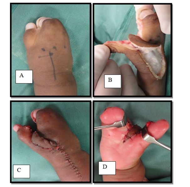
Operative technique (Figure 2). A linear palmar incision through the first web to the dorsum of the thumb was made to release the web space. All fibrous bands, contracted fascia and muscles between the first and second metacarpal bones were excised to achieve maximum web widening and deepening, allowing thumb abduction.

Figure 2 Drawing of the flap on the dorsum of the hand (A). A DMCA flap extend proximally was raised, rotated and then mobilized to cover the webspace (B). The flap is inset demonstrating minimal bulk. The donor site was closed primarily. The flap is extended onto the palm (D).
The axis of the flap was the midline between adjacent metacarpals. The pivotal point is 1.5 cm proximal to the leading edge of the theoretically second web space. To measure the length of the flap, we calculated the distance from that point of reference to the distal edge of the area to be covered, regardless of how far it goes beyond the web, in the palm. The dissection plane of the skin paddle locates between the fascia and the extensor paratenon. After verifying the flap perfusion and viability, we proceeded to its rotation 90 degrees in order to cover the first web space. The skin defect over the donor site could be closed directly. Complex syndactyly of the ulnar digits was released 9 months after surgery.
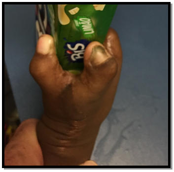
In this case, the position of maximum abduction was maintained with post-operative splint and kept in full abduction at night with a thermoplastic splint for 6 months after surgery.Initially, when he started to use, a useful trick to prevent from escaping the splint included the use of cobanover on it. The donor site heals without complication. Passive stretching, exercise programs at home were also performed. Complex syndactyly of the ulnar digits was released 9 months after surgery. After two years of the procedure, the function of pinch it was reached (Figure 3).

Figure 3 The well-settled appearance of the DMCA flap. Photography after two years, satisfactory function.
After seven years of follow up, the improvement of function and adaptation of the hand was satisfactory. Hand therapy was present during the follow-up with strategies to prevent scar retraction and stimulate the pinch and grasping and others movements with playful techniques (Figure 4). The goal is alwayshelping the child andtheir family achieve goals of independence and improved self-esteem. In the last evaluation, the patient was able to hold, interact, use and exchange objects between the hands (Figure 5).
The presence of an appropriate thumb-index commissure is a critical prerequisite in the functions of pinch and grasping the hand.5
The space of the commissure is shaped like a tetrahedron, with the distal skin forming a curve that extends from the metacarpophalangeal joint of the second finger to the area immediately distal to the metacarpophalangeal joint of the thumb.6
In the treatment of narrow or syndactylism of the first web, we must consider the therapeutic possibilities and choose the one that provides the best result with the lowest possible morbidity.7 On mild and moderate retractions, zetaplasty techniques are insufficient coverage options.8,9
In the other way, rotation flaps of dorsum can be used, as long as the skin is intact, however they haven't a large arc of rotation and length, and sometimes, skin grafting could be necessary for the donor area.10,11
Despite being indicated and available in severe retractions, free flaps seem to be very complex solutions, as well as remote pedicled flaps on the forearm and their disadvantages.12
The possibility of using a pedicled flap from the dorsal region of the hand showed very attractive.9 It is a flap close to the receiving area, thin, flexible, with a simple dissection technique, with wide arc of rotation and minimal donor area morbidity.13
And why was it possible to raise a longer flap? Omokawa.14 identified axial cutaneous arterial networks between the cutaneous branches of the dorsal metacarpal arteries and the cutaneous branches of the dorsal carpal arch. The cutaneous branches of the dorsal carpal arch anastomose with the cutaneous branches of the posterior interosseous artery (PIA) of the distal forearm, thus forming an additional axial cutaneous arterial network. According to these anatomic studies, a reverse dorsal hand flap can be safely modified to extend proximally to the dorsum of the distal forearm. According to these anatomical studies, the reverse flap of the dorsal metacarpal artery can be safely extended to the proximal region of the wrist, towards the forearm.15
Yours versatility allowed, in a simple and safe way, a good quality coverage for the first commissure, and when necessary, the same flap, longer, made it possible to cover cases of more extensive retractions, which affected the palm of the hand.
The purpose of presenting this case is to demonstrate the applicability of the dorsal metacarpal artery perforator flap in a specific case with thumb-index complex syndactyly, since it is a thin, pliable flap that is simple to raise, has wide arc of flap rotation and minimal donor-site morbidity, and can reliably cover soft-tissue defects up to the first web space, extended into the palm. It's important to remember that the satisfactory outcomes are attached with not only the surgery but also a good follow-up and focus on rehabilitation function.
This flap can be considered an alternative in dealing with this situation, moreover it could also be used in all cases of web retractions, either by burns, traumatic or congenital causes.
None.
No potential conflict of interest relevant to this article was reported.

©2020 Ribak, et al. This is an open access article distributed under the terms of the, which permits unrestricted use, distribution, and build upon your work non-commercially.