eISSN: 2574-9838


Case Report Volume 5 Issue 4
Plastic, Reconstructive Surgery and Burns Unit 37 Military Hospital, Ghana
Correspondence: Kwesi Okumanin Nsaful, Plastic, Reconstructive Surgery and Burns Unit 37 Military Hospital, Accra, Ghana
Received: July 05, 2020 | Published: July 2, 2020
Citation: Nsaful KO, Gentil S. Use of homodigital reverse island flaps for digital reconstruction – the perspective of a reconstructive surgery unit in Accra, Ghana. Int Phys Med Rehab J. 2020;5(4):136?139. DOI: 10.15406/ipmrj.2020.05.00246
Introduction: Several Local flaps can be used for the reconstruction of digital soft tissue defects with exposure of tendons and/or phalanges. The homodigital flap is a versatile option. This article discusses the use of the homodigital reverse vascular island flap-a regional, axial-patterned skin flap in the reconstruction of distal digital defects.
Methods: 6patients with a soft-tissue defect at the distal part of the finger were treated by homodigital island flaps for reconstruction. We evaluated the active range of motion of the involved finger, and the patient's satisfaction with the appearance of the finger after reconstruction.
Results: All patients admitted to a good functional outcome. The donor site morbidity was minimal. The take of the split-thickness skin graft to the flap donor site was generally good. However one patient complained of numbness of the finger over the donor site.
Conclusion: The Homodigital flap is a handy multipurpose flap that can be used. Notwithstanding its limitation, it is easy to raise and it can be used to a variety of defects.
Keywords: digital defects, local flaps, homodigital flap, active range of motion, split thickness skin graft
Digital injuries with soft tissue defects as well as with bone injury are a common presentation to the Trauma Emergency Room. Several Local flaps can be used for the reconstruction of digital soft tissue defects with exposure of tendons and/or phalanges.1,2 All these flaps also meant to be employed to cover tendons to minimizing formation of adhesions to allow gliding of tissue over the tendon.
The aim of digital soft tissue defect reconstruction with the use of local flaps is to obtain wound coverage and a painless scar, to preserve adequate and functional length of the digit, to preserve the sensibility of the fingertip, and the interphalangeal joint motion.3
Some of the options available to the reconstructive surgeon include the Anterior triangular flaps (Atasoy/V-Y flap)4 heterodigital neurovascular island flaps,5–8 the cross-finger flaps are useful alternatives.9–12
Additionally, free tissue transfers can be used in selected patients. Free flaps often employed for this purpose including groin skin flaps, groin osteocutaneous flaps, groin chimeric flaps, second dorsal metacarpal artery flaps, and partial toe flaps for digital reconstruction.13
In spite of all these options, the homodigital flap is a useful, helpful, and easy to raise flap which enriches the armamentarium of the reconstructive surgeon. One of the many advantages of the homodigital flap is the fact that it is a single-staged procedure that is confined to the injured digit.
Weeks and Wray in 1973 described the homodigital island flap that is based on the volar blood supply of the fingers, either the radial or ulnar digital artery and its venae comitantes.14 The flap can be harvested on a proximal (antegrade) or a distal (retrograde) pedicle.15 Proximally based flaps are used to cover more proximal defects, whereas reverse pedicle digital island flap described by Lai and colleagues16 are used to cover more distal defects over PIP and DIP joints.
The main disadvantages of the homodigital flap, the fact that it is not suitable for patients with peripheral vascular disease. Thus it is recommended that a digital Allen’s test be performed when considering the option of the homodigital flap.17 Another significant disadvantage is the risk of damage of the vascular pedicle as well as decrease of sensation over the donor site.
For the reverse pedicle digital island flap, the flap is outlined on the lateral border of the base of the affected digit. By reverse planning an estimation of the size of the flap required is made. As a rule the flap must be designed to be slightly larger than the actual defect to account for primary skin contraction. Dissection is carried out from proximal to distal until enough length of the pedicle is obtained, which usually corresponds the level of the DIP joint. During dissection- which is best done loupe magnification - effort must be made to identify the digital nerve. The nerve is then gently separated from the vascular pedicle and the digital vessel is ligated proximally. The elevated flap is then rotated into the defect and sutured loosely to avoid compression of the pedicle. One also ought to ensure that the rotation of the flap doesn’t result in kinking of the vascular pedicle. A Split thickness skin graft (STSG), harvested from the ulnar aspect of the dorsum of the hand, is usually used to close the flap donor site.
The flap is outlined over the lateral aspect of the donor proximal phalanx according to the size and shape of the defect. The junction of the dorsal and volar skin in the lateral aspect of the digit forms a line sometimes referred to as the “ Lai’s line”.16 Surgery is carried out using a digital block together with local anaesthesia to the skin graft site or a wrist block with the aid of tourniquet control and loupe magnification. General anaesthesia was only employed in the paediatric population. The skin incision is started from the proximal site of the flap. A lazy- S skin incision over the lateral surface of the digit is continued distally after division of the: proximal end of the digital artery. A thin cuff of tissue is left around the vascular pedicle and the latter should be dissected to the level at which the flap can easily reach the defect. Following release of the tourniquet haemostasis is secured and the flap is inset with interrupted 4/0 nylon sutures. A split thickness skin graft (STSG) from the dorsal ulnar aspect of the hand was applied on the flap donor defect and secured with a tie over bolster dressing. The involved digits are temporarily immobilized with a wooden spatula. The hand is elevated postoperatively to minimize venous congestion of the flap.
Clinical cases
(Table 1) 5 homodigital flaps were carried out between June 2019 and December 2019.
|
srl |
age |
Sex |
Reason for flap |
Closure of donor site |
Complications |
|
1 |
45 |
F |
Cover for Allen type 2 Fingertip injury Right Index Finger |
SSG |
|
|
2 |
9 |
F |
Flexion contracture Right Thumb |
10 closure |
|
|
3 |
41 |
M |
Shortened Right Middle Finger |
SSG |
|
|
4 |
23 |
F |
Fibroma Dorsum Right Index Finger |
SSG |
|
|
5 |
18 |
F |
Contracture Left Little Finger |
SSG |
Numbness of the finger over the donor site |
|
6 |
27 |
M |
Contracture Left Ring Finger |
SSG |
|
Table 1 Demographic and clinical characteristics of study participants
Description of outcome results and complications is reported in Table 1. Decrease of sensation over the donor site was a complaint made by 1 patient post-operatively.
Case 1 (Srl4)
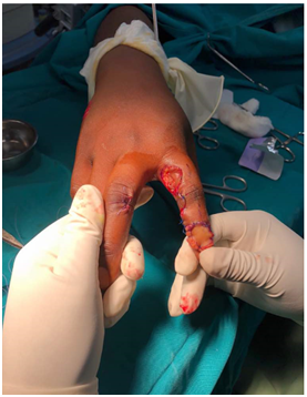
A 23 year old lady with a 6 month history of a soft tissue swelling over the dorsum of distal phalanx of the right index finger. Physical examination and X- rays done gave an impression of a Fibroma. The plan was to have an excision biopsy done and to cover the defect with a homodigital flap. By reverse planning the flap was designed and marking done to raise the flap (Figure 1a). The Fibroma was excised and the flap was raised (Figure 1b) and secured to the excision site, raised flaps were closed and the flap donor site was covered with STSG (Figure 1c).Healing was uneventful (Figure 1d).

Figure 1c Flap secured at the excision site, raised flaps sutured back into position, the donor site covered with STSG.
Case 2 (Srl3)
Case of a 41 year old male with a shortened right middle finger (Figure 2a). This is said to have happened when he sustained an Allen Level 4 traumatic amputation on the right middle finger. Finger lengthening was achieved by employing the homodigital flap (Figure 2b).
Case 3 (Srl2)
Nine year old girl presented with a seven year history of post burn contracture. Flap design markings were done.
(Figure 3a). An H incision was used to release the contracture and the post release wound was covered with the homogital flap. Due to skin laxity, the flap donor site was closed primarily (Figure 3b).
All the patients were generally happy with the outcome post-surgery. The lateral zig-zag incision or the lazy-S scar and the grafted donor defect were hidden well in the shadow of the inter digital space except for some hyper pigmentation of the grafted areas in some cases. Paraesthesia was noted in the ipsilateral dorsal skin in 1 patient. This symptom, however, was negligible in patients’ daily life according to his own statements. None of the patients had any stiffness in any of the joints of the fingers. They were able to achieve normal function and range of movement in the fingers on the average by 3 months post-operatively.
Even though there are several complex methods for correcting finger injuries, the Homodigital flapis a handy multipurpose flap that can be used. Notwithstanding its limitation, it is easy to raise and it can be used to a variety of defects.
None.
The authors declare no conflicts of interest.

©2020 Nsaful, et al. This is an open access article distributed under the terms of the, which permits unrestricted use, distribution, and build upon your work non-commercially.