eISSN: 2574-9838


Research Article Volume 2 Issue 4
2Department of Biomedical Engineering, University Camilo Castelo Branco, Brazil
2Electro-Electronic Systems, Sao Jose dos Campos, Brazil
3Department of Applied Physiology and Kinesiology, Sao Jose dos Campos, Brazil
Correspondence: Joao Paulo Alves do Couto, Department of Biomedical Engineering, University Camilo Castelo Branco: Parque Tecnologico de Sao Jose dos Campos, Brazil, Tel 557381531743, Tel 557381531743
Received: September 01, 2017 | Published: December 15, 2017
Citation: Couto JPA, Crespim L, Neto OP. The influence of different visual stimuli and head movement on the control of postural sway. Int Phys Med Rehab J. 2017;2(4):93-97. DOI: 10.15406/ipmrj.2017.02.00057
This study examined how different visual stimuli can alter the center of pressure’s sway pattern of healthy individuals in the standing position and if the changes in sway pattern due to visual stimuli occur because of head movements or eye movements. We measured postural sway in 25 healthy individuals. The visual stimulus was displayed at eye level. The individuals remained standing, on a pressure platform in two situations, one with use of the Philadelphia neck collar and the other without. Each individual experienced the 13 combinations of visual movements with six combinations using the neck collar and seven combinations without. Were used, three frequencies of horizontal movements, three frequencies of depth movements and one with the sphere motionless. The results showed: Increase in sway of the anteroposterior axis when not using the neck collar; The type of visual stimulus affected specific frequency bands of body sway in the anteroposterior axis; Stabilometric differences between genders; and Increase in the total average speed of the center of pressure for the lateral visual stimulus without the use of the neck collar.
Keywords: postural sway, visual stimulus, eyes movements, head movements, visual perception
Visual, proprioceptive and vestibular systems clearly contribute to postural control, as several studies have shown that visual1 proprioceptive2 or vestibular3 stimulation evokes and controls body sway. Each sensory system detects an "error", indicating a deviation in the body’s orientation from a reference position. The visual system is considered to be the most complex4 of the sensory systems and has a central importance.5 It’s operation involves structures and mechanisms for obtaining information, which are obtained through the refraction of light from the surfaces of objects, plants, animals, etc. Visual conflicts can have powerful effects on balance. Visual movements in the environment can cause postural changes and imbalance, as well as motion sickness in healthy adults.5 According to Ehrenfried7 a problem with the results of many studies in this area has been the variability and the apparent inconsistency in responses. A hypothesis exists that there may be a functional relationship between body sway and visually guided eye movements. This is because the accuracy of such movements can be influenced by small changes in body sway. On the other hand, eye movements that are not visually guided are not subject to being facilitated or interrupted by small changes in body sway.8
The researchers that have addressed the relationship between eye movements and body sway tend to predict that sway should increase during eye movement, in relation to the swaying that is present when the eyes are not moving. There are two reasons for this prediction. One is based on the concept of between-task competition for limited central processing resources.9–10 From this point of view, cognitive resources dedicated to the control of eye movements would not be available for the control of posture. Therefore, we can expect an increase in body sway when people use eye movements, for example, when following the motion of a visible target.11
The second reasons is based that the rotation of the head (tracking of the visual stimulus) is a problem in the assessment of body sway. This occurs because three reasons: it becomes more difficult to understand the role of eye movement, by itself, in postural control; it tends to present non-postural head movement which complicates the interpretation of postural measurements; and the rotation of the head can bring into play the so-called vestibulo-ocular reflex12 which could complicate the interpretation of both visual and postural behavior.13 This study aims to better understand how different visual stimuli can alter the center of pressure’s sway pattern of healthy individuals in the standing position. In addition, it seeks to understand whether changes in sway pattern due to visual stimuli occur as a result of head movements or not.
This research protocol followed the ethical principles of the Brazilian Guidelines for Research-CAAE(Protocol n° 01092112.5.0000.5062).
Subjects
25 healthy individuals, of whom 10 women and 15 men with ages ranging between 18 and 38 years old, who provided their informed consent to participate in the study.
Visual stimulus
Display of the visual stimulus was rendered by using an IPAD screen, displaying a video imaging software generating a green sphere against a black background. This software was developed especially for this study. The image consisted of a sphere with a radius of 1.25cm on a screen 14.9cm in height and 19.8cm wide. The ball made horizontal movements (Figure 1) as well as movements of depth (Figure 2) as selected by the researcher. The speed of oscillation of the sphere for both the vertical and horizontal movements were also selected by the researcher and varied between 0.5Hz, 1Hz, and 2Hz.
The image was displayed at eye level at a distance of 65cm from the eyes of each individual involved in the study. The individuals remained standing with their feet in parallel and separated, as aligned with their shoulders, on a pressure platform (Splate 2.52) in two situations, one with use of the Philadelphia neck collar (Figure 3) and the other without the use of the neck collar (Figure 4). The neck collar weighed 300 g and was placed (S, M, and L) on the participants according to the patient’s height. This approach was utilized, considering that the aim was to obtain postural control for tasks in a manner in which it would be sensitive to disturbances, while not being at risk of the individual taking a step or making hip movements in order to preserve balance. Ehrenfried et al.7 The lab environment was free from noises that could distract the attention of the individuals from the IPAD’S screen. The individuals remained in the room with the researcher and no other individual being present.
Postural sway
The movements of the foot’s center of pressure (FCOP) in the frontal and sagittal planes were measured and translated by the S-Plate pressure platform and Software Version 1.10. The S-Plate pressure platform has size (Length/Width) 610x580mm, active surface 400x400mm, resistive sensors type, sensors number-1600. This product was tested and approved for DEKRA Certification GmbH (Identification Number 0124) approved the Quality System. The signals were collected for 20s at 5Hz.
Task
Each individual experienced the 13 combinations of visual movements with six combinations using the neck collar and seven combinations without use of the neck collar. Were used in the study, three frequencies (0.5Hz, 1Hz, and 2Hz) of horizontal movements (Right, Left), three frequencies (0.5Hz, 1Hz, and 2Hz) of depth movements (anterior, posterior) and one with the sphere motionless. The selection of the sequence in which the visual movements were exposed to the individuals was randomized. During the task, the participants were not allowed to perform support if necessary.
Variables
Based on the center of pressure displacement data, variables were calculated in the time domain and in the frequency domain. The variables calculated in the time domain were: standard deviation of the displacement in the anteroposterior direction (SDAP); standard deviation of the displacement in the latero-lateral direction (SDLL); root mean square of the displacement in the anteroposterior direction (RMSAP); root mean square of the displacement in the latero-lateral direction (RMSLL);and total average speed (TAS). The variables calculated in the frequency domain were: Integral of power in different frequency bands of the anteroposterior displacement (y-Power) and integral of power in different frequency bands of the latero-lateral displacement (x-Power). The spectra of the center of pressure displacements were obtained using the fast Fourier transform (FFT). The frequency bands chosen were: 0 to.7Hz, 0.7 to 1.4Hz, 1.4 to 3Hz, and 3 to 5Hz. Frequency bands were chosen considering the frequencies of visual stimulation. Fourier transforms were calculated considering a 200 ms window size and 50 ms overlap.
Statistics
Mixed analyses of variance (ANOVA) of repeated measures with several factors were used to determine possible main effects and significant interactions. For analysis of the variables obtained in the time domain a mixed ANOVA with 2 inter-subject factors was used: frequency of oscillation of the virtual sphere which varied between 0.5, 1Hz, and 2Hz (Frequency factor) and type of movement of the virtual sphere which varied between side to side movements and the simulation of movements in and out of the screen (Type factor); as well as two intra-subject factors: use or nonuse of a neck collar (Neck factor) and the participant's gender (Gender factor). In order to analyze the variables of the frequency domain an additional inter-subject factor was considered: the frequency band analyzed (Band factor).
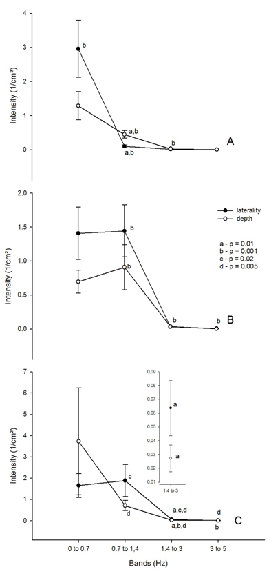
The results suggest that the neck collar significantly affected (F6.144=3.245; p=0.005) specific frequency bands of body sway of the participants in the anteroposterior direction differently for the three frequencies of visual stimuli used, regardless of the stimulus type (laterality or depth). The participants that did not use a neck collar, when exposed to a visual stimulus at a frequency of 0.5Hz showed greater sway in the anteroposterior axis in the bands of 0 to 0.7Hz, 0.7 to 1.4Hz (p=0.002), and 1.4 to 3Hz (p=0.01) when compared to those who did wear the neck collar (Figure 5A). When exposed to a visual stimulus at frequency of 1Hz, the participants that did not use the neck, showed greater sway in the anteroposterior direction in the bands of 0 to 0.7Hz (p=0.05), when compared to those who did wear the neck collar (Figure 5B). Finally, participants when exposed to a visual stimulus with a frequency of 2Hz showed no significant differences between the behavior of the bands of frequency of body sway in the anteroposterior axis, whether the neck collar was used or not (Figure 5C).

The results also showed that the type of visual stimulus (laterality or depth) significantly affected (F6.138=2.750; p=0.04) specific frequency bands of body sway of the participants in the anteroposterior direction differently for the three frequencies of visual stimulus used, regardless of using the neck collar or not (Figure 6). The participants when exposed to a visual stimulus at a frequency of 0.5Hz (Figure 6A) while receiving a visual stimulus of depth showed greater sway in the anteroposterior axis in the bands of 0.7 to 1.4Hz, when compared to when they received the lateral visual stimulus (p=0.01). When exposed to a visual stimulus at a frequency of 2Hz (Figure 6C) the individuals receiving the depth visual stimulus presented greater sway in the frequency band of 0 to 0.7Hz, whereas in the band of 1.4 to 3Hz sway in the anteroposterior axis was greater for the individuals who received the lateral stimulus (p=0.01).

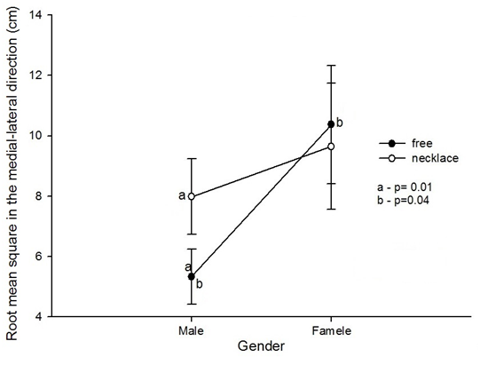
In addition, the gender of the participants as well as whether or not the neck collar was used significantly affected (F1.23=5.453, p=0.02) the root mean square in the medial-lateral direction (Figure 7). Regarding the gender of the participants, the root mean square of the medial-lateral direction of the male participants that did not use the neck collar was lower than that of the female participants under the same condition (p=0.04). Whereas if we consider use or nonuse of the neck collar, the results indicate that the male participants who did not use the neck collar had lower root mean square values in the medial-lateral direction when compared to those that did use the neck collar (p=0.01) (Figure 7).

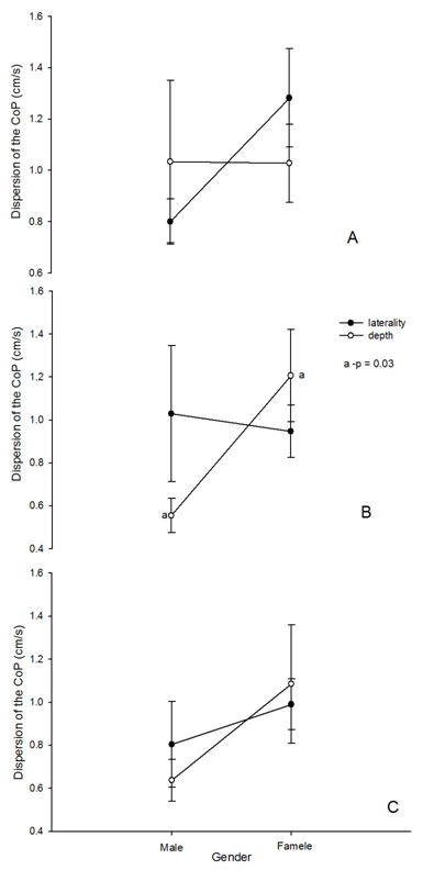
The results also indicate that the gender of the participants and the type of visual stimulus significantly affected (F2.46=3.763 p=0.03) the dispersion of the COP displacement from the mean position in the anteroposterior direction, in a different manner for the three visual stimulus frequencies utilized (Figure 8). When the participants were exposed to a visual stimulus at a frequency of 0.5Hz they showed no significant differences between the type of stimulus and the gender of the participants (Figure 8A).
Female participants, on the other hand, when exposed to a visual stimulus at a frequency of 1Hz of depth had a greater dispersion of the COP displacement from the mean position in the anteroposterior direction when compared with the male participants (p=0.03) (Figure 8B). Finally, when the participants were exposed to a visual stimulus at a frequency of 2Hz they showed no significant differences between the type of stimulus and the gender of the participants (Figure 8C).

Finally, the type of visual stimulus (laterality and depth) as well as the use of the neck collar significantly affected (F1.23=5.89, p=0.02) the total average speed of the center of pressure (Figure 9). When participants were exposed to the lateral visual stimulus the total average speed of the center of pressure was higher without the use of the neck collar (p=0.02).
Our study observed how the central visual stimuli of depth and laterality, at three different frequencies (0.5Hz, 1Hz, and 3Hz), as well as head motion influence body sway. The results showed the following findings: Visual stimuli caused Increase in sway of the anteroposterior axis when subject were not using the neck collar; The type of visual stimulus (laterality or depth) affected specific frequency bands of body sway in the anteroposterior axis; Stabilometric differences between genders in dispersion of the COP displacement from the mean position in the anteroposterior direction; The lateral visual stimulus caused an increase in the total average speed (TAS) of the center of pressure when the subjects were not using the neck collar. In our study we tried to verify whether there is an influence of rapid movement of the head on body sway when performing an eye tracking task. For this purpose we placed Philadelphia-type neck collars on the participants in order to restrict the tracking movement of the head. The participants were exposed to the same stimuli under two conditions: without the use of the neck collar and with neck collar use.
We can see in our results (Figure 5) that the participants that did not use the neck collar showed greater sway in the anteroposterior axis in the bands of 0 to 0.7Hz, 0.7 to 1.4Hz and 1.4 to 3Hz, when compared to those who did wear the neck collar, especially when the visual stimulus was moving at a frequency of 1Hz or less. Ojima et al.14 have shown that the low-frequency of body sway is determined mainly by the movement of the head. Thus, body sway has a component of low frequency dictated by the movement of the head, and a high-frequency component that is caused by other parts of the body. At low frequencies, however, the movement of the head provides greater power, and at higher frequencies body sway prevails. The fact that body sway is clearly greater than the movement of the head in the high frequency range implies that the high frequency components are related to some other sources.
When participants were submitted to depth stimulation they reported that they felt as if their body was being pushed and pulled in the anteroposterior direction; these reports were similar to those in the study by Kuno et al.15 where six of the 10 participants of their study reported that they felt they were pushed by the pattern of depth optokinetic stimulus (DOKS) when the sample approached them. This caused them to move back. When the DOKS model (stimulus) moved away from them, they reported that it made them feel like they were being pulled forward. However, this account was not as consistent with the stabilometric data given the fact that the participants who underwent the depth stimulation swayed in the anteroposterior axis as much as or less than the participants who were subjected to the lateral stimulus. When the participants were exposed to depth and lateral visual stimuli at 0.5Hz, the anteroposterior sway with static difference occurred in the band from 0.7 to 1.4 for those who received the depth stimulus. However, when the frequency of the visual stimulus was 2Hz the frequency bands of anteroposterior sway that showed statistical differences were 0.7 to 1.4Hz and 1.4 to 3Hz for those that received the lateral stimulus. Kunkel et al.16 suggest that the frequency of spatial stimulation turns out to be a decisive quality of visual stimuli for the stabilization of posture, since spatial frequencies with the lowest perceptual boundaries correspond to the frequencies with the best postural performance. The authors also go on to say that the more complex geometry of stimuli which move in the sagittal direction may involve higher visual areas such as the radial superior temporal gyrus which may contribute to the differences between anteroposterior and lateral sway in the stabilization of posture.
Another point observed in our study was that men and women had different results when evaluating stabilometric parameters. Women had a root mean square in the medial-lateral direction (RMSML) greater than that of men and the Dispersion of displacement of the COP from the middle position in the anteroposterior direction (SDAP) when visually stimulated by depth was at a frequency of 1Hz. Chiari et al.17 explain that most of the gender effects on stabilometric parameters are actually the result of differences in biomechanical properties rather than differences in the posture control system. In their studies the authors also explain that the regression analysis showed that the parameters which quantify sway (such as the influence of the path, average speed or time interval) are strongly dependent on "height" (and partly on "weight") and that "height" must be considered as a major anthropometric determinant between genders.
We also found that the participants who did not use the neck collar and received the lateral visual stimulus had a TAS greater than those who used the neck collar (Figure 9). This is explained by the fact that body sway is visually induced by the increase in speed of the visual stimulus up to a certain level. This level corresponds to the optokinetic limitations on the optokinetic nystagmus15 According to Stoffregen et al.8 there may be a functional relationship between body sway and visually guided eye movements. This is because the accuracy of such movements can be influenced by small changes in body sway.
In summary, the data presented herein, on the relationship between the different visual stimuli at different frequencies and head movement showed that an increase in TAS, in the lateral direction, for the participants who did not use the neck collar occurred due to visually guided eye movements, whereas the increase in body sway that occurred in the anteroposterior direction is mainly due to head movement. When gender differences are analyzed it can be observed that biomechanical factors, especially height, also have an influence on postural sway. This article can serve as a basis for the construction of knowledge and the starting point for new research. However, further research in this direction is needed.
Supported by Fapesp Processo 2012/09400-9 to Osmar Pinto Neto.
The author declares no conflict of interest.

©2017 Couto, et al. This is an open access article distributed under the terms of the, which permits unrestricted use, distribution, and build upon your work non-commercially.