eISSN: 2574-9838


Short Communication Volume 7 Issue 3
Department of Engineering, Toyota Technological Institute, Japan
Correspondence: Masami Okamoto, Advanced Polymeric Nanostructured Materials Engineering, Graduate School of Engineering, Toyota Technological Institute, 2-12-1 Hisakata, Tempaku, Nagoya 468 8511, Japan, Tel +81 52 809 1861
Received: November 23, 2022 | Published: December 2, 2022
Citation: Sasaki R, Ohta R, Okamoto M. Stemness of breast cancer cells incubated on viscoelastic gel substrates. Int Phys Med Rehab J. 2022;7(3):136-137. DOI: 10.15406/ipmrj.2022.07.00320
Acrylamide copolymer-based gel substrates with different viscoelasticity were employed to evaluate the viscoelasticity effect on the direct relation among cancer stemness and mesenchymal properties with induction of epithelial–mesenchymal transition (EMT) of human breast adenocarcinoma (MCF-7) cells in both normoxia and hypoxia. The softer gel substrate produced a large amount of surface molecule of cancer stem cells (CSC) marker CD44. In contrast, for the stem cell biomarker CD133 expression, their coefficient of damping (tan)-dependent manner was not contributed by EMT phenomenon and was an independent from acquisition of the EMT. The substrate damping as potential physical parameter emerged the important linkage to cancer stemness and EMT induction.
Keywords: viscoelastic gel substrates, cancer cells, epithelial-mesenchymal transition, gene expression,
cancer stem cells
CSC, cancer stem cells; EMT, epithelial–mesenchymal transition; DMEM, dulbecco’s modified eagle’s medium
Cancer ranks as the leading causes of mortality in Japan.1 A recently proposed hypothesis suggests that cancer stem cells (CSCs) and epithelial–mesenchymal transition (EMT) play a pivotal role in cancer metastasis, recurrence and drug resistance.2 A few CSCs do self-renew and produce a large number of heterogeneous and highly proliferative cancer cells to form primary tumors. Thus, CSCs survive and cause cancer recurrence. Recent studies showed that inducible factors of EMT not only induce EMT, but also enhance CSC features of cancer cells.2 These findings suggest that EMT is closely related to cancer progression.
Our previous study showed that scaffold-induced EMT of mammary carcinoma cells could be established in vitro by using a polymer cell culture scaffold with a three-dimensional fibrous porous structure.3 In addition, the relationship between EMT and the motility of mammary carcinoma cells under both hypoxia and normoxia was studied using polymeric gel substrates with different viscoelasticity, which mimic in vivo micro- environment.4
In this study, we aimed at examining the effect of viscoelasticity of the substrate on the direct relation among cancer stemness and mesenchymal properties with induction of EMT in both normoxia and hypoxia.
Experimental
Cell culture: Human breast adenocarcinoma cell line, MCF-7 (ATCC) were cultured in high glucose Dulbecco’s modified Eagle’s medium (DMEM) (Nacalai Tesque, Japan) supplemented with 10% (v/v) FBS, grown at 37°C under 5% CO2 atmosphere and 95% relative humidity (normoxia) or hypoxic condition (94% N2, 5% CO2, and 1% O2) at 37°C.
CSC marker expression
MCF-7 cells were seeded at a density of 5.0×10³ cells/cm2 on eight different substrates, and cultured for 7 days. The substrates were co-polymerized gels (P(AAm-co-ACA)) (AC)4 made from copolymers of Acrylamide (AAm) and N-acryloyl-6-aminocaproicacid (ACA) made into six different hardness, and coated with Cellmatrix Type I-C (Nitta Gelatin). In addition cell culture plates (TCP: control) and collagen-coat coated with TCP and Cellmatrix Type I-C were used. After culturing, the expression levels of CD133 and CD44 were measured by fluorescence excitation preparative assay using a flow cytometer (Attune NXT Acoustic Focusing Cytometer, Thermo Fisher Sci. Flow Jo (ver 10) was used as analysis software.
Gene expression analysis
The expression level of HIF-1α, TGF-β, vimentin, CDH2, CDH1, SNAI2, ZEB1 were evaluated by RT-PCR.4 Statistical analysis was performed using Tukey–Kramer method.
CSC marker expression
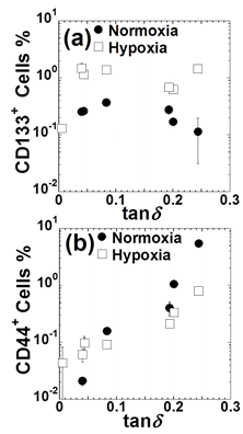
The expression rates of CD44 and CD133 in AC gels as a function of viscoelasticity are shown in Figure 1. The CD133+ levels increased the loss factor (tanδ) of AC gels to around 0.09 under both oxygen conditions and then decreased. The CD44+ levels increased with the increase in tanδ under both O2 conditions, indicating for the first time that the expression of CD133 and CD44 is not related to each other under different O2 conditions in MCF-7 cells. CD44 is known to bind mainly to hyaluronan and is involved in the maintenance of CSCs. On the other hand, the expression of CD133 has been found to correlate with poor prognosis.5 Since each transmembrane protein contributes to different functions, it is suggested that the dependence on tanδ is also different.

Figure 1 Flow cytometry analysis of (a) CD133, (b) CD44, expression level of MCF-7 cells incubated on different viscoelastic gel substrates and collagen-coated TCP under both oxygen concentration conditions for 7days. Data were expressed as mean ± SD (n=3)..
Gene expression
MCF-7 cells cultured in AC gels with tanδ= 0.244 (AC-soft) and tanδ= 0.195 (AC-mid) showed significant changes in vimentin expression under both oxygen concentration conditions. The expression of vimentin under hypoxia was significantly increased by 720-fold in AC-soft and 360-fold in AC-mid gels compared to AC gels with tanδ = 0.0437 (AC-stiff). The expression of CDH2 was similar to that of vimentin, and the expression of CDH2 was 440-fold higher in AC-soft than in AC-stiff under hypoxia.
This work was supported by the TTI Grant (Special Research Project: FY2021).
Contributed to the initiating idea and performed most of the experiments: Ohta Y, Sasaki R, and Okamoto M. Supervised research: Okamoto M. The manuscript was written through contributions of all authors.
Author declares that there is no conflicts of interest.

©2022 Sasaki, et al. This is an open access article distributed under the terms of the, which permits unrestricted use, distribution, and build upon your work non-commercially.