eISSN: 2574-9838


Case Report Volume 2 Issue 3
Department of echocardiography, Instituto de Cardiología, Tucumán, Argentina
Correspondence: Jorge Tazar, Department of echocardiography, Instituto de Cardiología, Tucumán, San Miguel de Tucumán, Tucumán, Argentina, Tel 5403814330389
Received: November 14, 2017 | Published: December 1, 2017
Citation: Tazar J, Palacio GM, Rojas NA, et al. Rare malformation of the left atrial appendage: report of a case. Int Phys Med Rehab J. 2017;2(3):59-60. DOI: 10.15406/ipmrj.2017.02.00050
In this report, we present a case of a rare malformation of the left atrial appendage. This finding was observed during the transesophageal echocardiogram performed on a patient diagnosed with supraventricular arrhythmia who was undergoing electrical cardioversion. This observed malformation of the left atrial appendage has not been previously described.
Keywords: left atrial appendage malformation, anatomy of the left atrial appendage
AF, atrial fibrillation; AVB, atrioventricular block; ECV, electric cardio version; L, left atrium; LAA, left atrial appendage; TEE, transesophageal echocardiography
In recent years, the anatomical study of the Left Atrial Appendage (LAA) has regained interest. Studies using cardiac nuclear magnetic resonance and multi slice computed tomography have described various morphologies of the LAA, namely: Cactus, chicken wing, windsock, and cauliflower.1 The hole that connects the LAA with the atrium has also been described. This route of entry is closely related to the arrival of the left superior pulmonary vein, the conduction system and the circumflex artery and can present different forms: Oval, circular, triangular or in drop.2 This report describes an LAA that manifests anatomical characteristics different from what is known.
A male patient, 66 years old, presents as risk factors a smoking habit that had ceased in the last 5 months. As a cardiovascular history, he had two previous admissions for supra ventricular arrhythmias, which were reversed in a timely manner with Electrical Cardioversion (ECV). The patient is admitted to the emergency room with symptoms of asthenia and adynamia of 72 hours of evolution. The 12 lead ECG showed an atrial flutter with a high degree of Atrioventricular Block (AVB), with a 5:1 conduction. It had a heart rate of 50 beats per minute (Figure 1). The patient received amiodarone 400mg./day. He was hospitalized in a coronary unit to try a new ECV. Blood pressure: 150/80mmHg. The exams of the other systems (respiratory, neurological, etc.) had no positive data. It is indicated to start anticoagulation with low molecular weight heparin, and a Transesophageal Echocardiogram (TEE) is requested to rule out cardio embolic source.
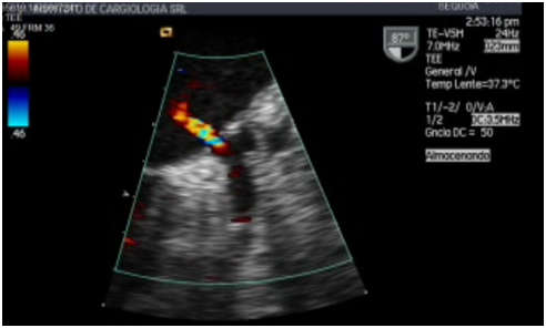
The patient was in the left lateral decubitus under deep sedation and with oropharyngeal anesthesia with xylocaine, TEE was performed with a multi planar probe. At the middle esophageal level, with an angle of 87degrees, it was possible to verify that the morphology of the LAA corresponded to the chicken wing type, with a size of 4.3 cm2 of area. It was noted that at the connection site between the LAA and the Left Atrium (LA) a ¨septum¨ that closure LAA. This structure showed no movement and when it was explored with color Doppler, it was found that it had a narrow perforation through which a high-speed bidirectional flow proceeded (Figure 2-5). In the interior of the appendage, there was no evidence of spontaneous contrast echo or thrombi.


The LAA is an embryological remnant of the primordial atrium and is formed by the absorption of the primordial pulmonary veins and their branches. It is a glove-like structure, generally located between the anterior and lateral wall of the LA and whose tip is directed anteriorly and superiorly, overlapping the left edge of the right ventricle outflow tract or the pulmonary trunk and the birth of the left or circumflex coronary artery. It is not uncommon to find the tip of the LAA in the lateral and posterior direction. However, in a small percentage of hearts, the tip of the LAA passes behind the arterial pedicle to settle on the transverse sinus of the pericardium.2
The main importance of LAA is that it is the site where thrombi are most frequently formed in patients with Atrial Fibrillation (AF); these thrombi carry the risk of subsequent embolism. Necropsy studies have described that in 98% of the cases thrombi formed in the atrium of patients with AF are found in the LAA.3 Taking into account these precedents, for several decades, they have been considered the closure of the LAA as a treatment in the prevention of stroke.4 In the beginning, the closure was performed surgically, either through a split or an exclusion. However, in recent years, the exclusion of the LAA has been performed with percutaneous closure devices. Currently, there are several models and there are still others in research.5 A report about a malformation at the level of the left atrial appendage is presented. There is no previous evidence that refers to this type of anatomy of the LAA and for our consideration it would be a rare congenital anomaly of the same. Characterizing the real clinical impact of this finding is difficult in light of the current knowledge.
The authors have the patient´s permission for the presentation of the case report.
None.
The authors do not present any conflict of interest.

©2017 Tazar, et al. This is an open access article distributed under the terms of the, which permits unrestricted use, distribution, and build upon your work non-commercially.