International Journal of
eISSN: 2574-9889


Mini Review Volume 11 Issue 2
 Devanshi Shah2
1M.S. OBGY, FICOG, Fellowship in Maternal-Fetal Medicine, UK
2M.S. OBGY, Diploma in fetal Medicine and Advanced gynaec ultrasound, Shree Krishna Hospital, Bhaikaka University, India
Correspondence: Nupur Shah, M.S. OBGY,FICOG, Fellowship in Maternal-Fetal Medicine, Fellowship in Medical Genetics, UK
Received: July 04, 2025 | Published: July 14, 2025
Citation: Shah N, Shah D. Prenatal ultrasound assessment of aortic isthmus and its biometry in Indian population. Pregnancy & Child Birth. 2025;11(2):36-37 DOI: 10.15406/ipcb.2025.11.00319
The aortic isthmus is the segment of the aorta between the origin of the left subclavian artery and the connection of ductus arteriosus to the descending aorta1, a narrow segment, which is important not only in context to diagnosis of coarctation of aorta and interrupted aortic arch, but also in fetal growth restriction and obstructive left ventricular outflow disorders (hypoplastic left heart, critical aortic stenosis) where the aortic isthmus flow reversal is noted. Fetal aortic isthmus measurement gives us an insight in achieving probable diagnosis of coaractation of aorta. Through our study, we aim to measure aortic isthmus in fetuses across gestational age of 16-40 weeks in Indian population, to determine the normative range and z scores for the gestation. This would aid in predicting any abnormality in context to coarctation and triage these fetuses for close follow-up postnatally to confirm the diagnosis.
A Longitudinal Observational study with 200 low risk fetuses with normal heart coming to the clinic of Author 1 at SKH hospital, from 1st August 2023 to 15th June 2024 between 16 – 40 weeks of gestation were taken. All the views were taken by FMF-UK certified operator using Voluson E8radiance, GE, Milwaukee, USA. The measurements were obtained with the heart in the optimal view with the cardiac apex nearer to the transducer. Aortic isthmus was measured in both the three-vessel trachea view and sagittal view as shown in Figure 1. The data was entered into the excel sheet. Descriptive and Regression analysis of the data was obtained using the STATA 14.2 software.
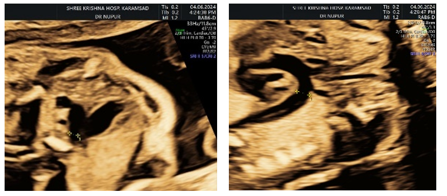
Figure 1 Aortic isthmal measurement in 3 vessel trachea view and the sagittal view.
Adequate sonographic measurements of aortic isthmus were obtained in fetuses with the gestational age between 17-24 weeks of gestation in both the sagittal and trans axial view.
The 5th and the 95th centile of the aortic isthmal diameter for gestational age between 17-24 weeks has been described in Table 1.
|
Gestational age |
5th centile |
95th centile |
|
17-17+6 |
1.23 |
1.4 |
|
18-18+6 |
1.3 |
1.73 |
|
19-19+6 |
1.35 |
2.02 |
|
20-20+6 |
1.52 |
2.11 |
|
21-21+6 |
1.6 |
2.2 |
|
22-22+6 |
1.72 |
2.48 |
|
23-23+6 |
1.7 |
2.79 |
|
24-24+6 |
2.1 |
2.85 |
Table 1 Gestation age versus 5th and 95th centiles of aortic isthmus diameters
The predicted mean value for both variables were obtained in all 200fetuses respectively as follows for 16-16 +6 (1.22), 17-17 +6 (1.33),18-18 +6(1.47),19-19 +6 (1.61),20-20 +6 ( 1.76) ,21-21 +6 (1.90) 22-22 +6 (2.04),23-23 +6(2.18), 24-24 +6 (2.32), 25-25 +6 (2.46), 26-26 +6 (2.60),27-27 +6 (2.74),28-28 +6 (2.88),29-29 +6 (3.02),30-30 +6 (3.16),31-31 +6 (3.30),32-32 +6 (3.44),33-33 +6 (3.58),34-34 +6 (3.72),35-35 +6 (3.86),36-36 +6 (4.00),37-37 +6 (4.14),38-38 +6 (4.28),39-39 +6 (4.42),40-40 +6 (4.56).
The mean value for the both variables showed a linear progressive trend with increasing gestational age (Figure 2). The aortic isthmus diameter was found to be increasing with advancing gestational age through statistical analysis(p<0.05). The regression analysis of aortic isthmal/ductal ratio to that of gestational age showed that it was close to a constant value of 1, regardless of gestational age. The mean was 0.90 with a CI of 0.79-0.92.
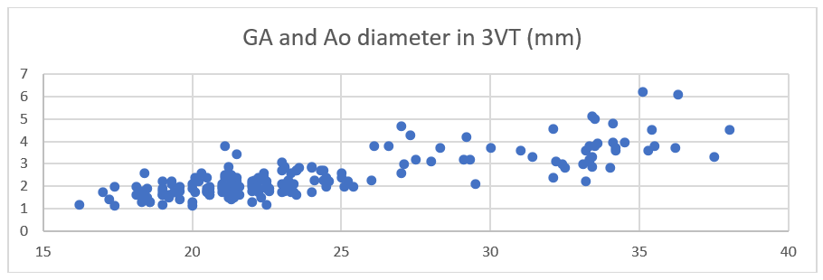
Figure 2 Scatter plot of aortic isthmus measurements btwn 16-40 weeks of gestation.
Aortic isthmus is the segment of the aorta between the origin of the left subclavian artery and the connection of ductus arteriosus to the descending aorta.Its measurement and generating of the normative data is important in predicting the abnormalities like coarctation. In this study, the aortic isthmyal diameter is measured in both views- transverse and sagittal, so as to include both the standard screening as well as extended fetal echocardiographic views.1
The mean and the lower and upper 95th centiles of aortic isthmus diameters measured by Achiron et al.,2 were similar to our study. However, Achiron et al.,2 only observed the aortic isthmal diameter in the sagittal view and not in the standard three vessel trachea view. Achiron et al.,2 also observed that the aortic isthmal diameter increases with advancing gestational age in accordance with our study.3–6
Nomiyana et al.,7 studied the aortic arch only in sagittal view only, where the morphology visualization can be limited. Although the sagittal view offers the visualization of the neck vessels the side-to-side comparison of the ductal and the aortic isthmus cannot be achieved in the sagittal view.8
Vigneshwaran et al.,9 studied the distal transverse aortic arch in 3 vessel trachea view in 7000 fetuses and obtained the z scores, and 5th and 95th centiles for gestational age. Their 5th and 95th centile of their study is comparable to our study.
Pasquini et al.,3 measured the aortic isthmus diameters in 204 fetuses in three vessel trachea view and 190 fetuses in sagittal view. They developed the Zscores and isthmal to ductal ratio against the gestational age. The regression analysis for the aortic isthmal to ductal ratio showed a constant value of 1 regardless of the gestational age which was also similar to our study. Pasquini et al.,3 also observed that the mean of the ratio was 0.99 with all ratio values expected to lie between 0.74 and 1.23.
Though the number of fetuses studies here was a limitation, the study provides a strong foundation for future larger studies and alsois one of its kind in the region.
Given the current lack of data for Indian population to the best of our knowledge, the normative data obtained through our study may be helpful. The study was conducted according to Declaration of Helsinki and consistent with GCP.
None.
The authors have no competing interests to declare.

©2025 Shah, et al. This is an open access article distributed under the terms of the, which permits unrestricted use, distribution, and build upon your work non-commercially.