International Journal of
eISSN: 2573-2889


Short Communication Volume 6 Issue 1
Department of Zoology, University of Allahabad, U.P, India
Correspondence: Sandeep K Malhotra, 16-A/1, Church Lane, Prayagraj- 211002, U.P, India, Tel +91 6389318380
Received: November 20, 2023 | Published: November 27, 2023
Citation: Yadav A, Malhotra SK. Critical add-on SEM observations on oral armature and life cycle of raphidascaridoid worm, indospinezia multispinatum Jaiswal and Malhotra (2017). Int J Mol Biol Open Access. 2023;6(1):59-62. DOI: 10.15406/ijmboa.2023.06.00153
Additional observations of taxonomic significance with unique life cycle details were recorded after the publication of specialized liver-inhabiting roundworms, Indospinezia multispinatum Jaiswal and Malhotra (2017) in the Gangetic gar-fish, Xenentodon cancila from Prayagraj riverine ecosystems. Typically strong musculature supporting triangularly placed one dorsal and two ventro-lateral lips has been observed and their characteristics recorded. The muscular complement that comprised each of the 3 lips, was 3-layered, each of which was stacked one over the other, around a medial furrow. The 2nd stage larva armed with cephalic tooth was the specific discovery which was extracted by Pepsin Digestion method, during compilation of various larval stages that made up complete life-cycle of the roundworm under study. Presumption based conclusions derived on larval stages in the life cycle of Goezia bangladeshi by Akther,1 et al led to erroneous interpretations and hence made the taxonomic position of Goezia bangladeshi circumspect unless appropriate larvae are studied. Therefore, Goezia bangladeshi is termed as species inquirendae pending complete description of its larvae.
Keywords: Indospinezia multispinatum and Xenentodon cancila, raphidascaridoid, tribe, life cycle, cephalic tooth, Goezia bangladeshi, species inquirendae, circumspect, trilayered lips
D, lid on oral aperture; F, furrow; K, knob like tip of spine; Lm, muscular rim of lip; MP, minute papillae; Sl, second muscular layer to strengthen lip; Tl, third muscular layer to strengthen lip
Extended investigations on the freshly collected roundworms, Indospinezia multispinatum parasitizing Gangetic garfish, Xenentodon cancila were conducted to study Scanning Electron Microscopic observations vide methodology followed in Jaiswal and Malhotra (2017), for hitherto unreported newer facts on the oral armature. Pepsin Digestion method Murill et al,2 was employed to extract 2nd and 3rd stage larvae along with earlier stages of larvae.
Single-toothed unidentate spines were recorded in the worms, the magnified view of which by SEM observations (Figure1a) exhibited additional 2-3 rings of minute spines over the main body of larger spines towards distal extremity. A small knob–like terminal tip (Figure 1b) on the distal end of the larger spines was also recorded as an additional feature. Additional observations by SEM emphatically illustrated the trilayered lips, that comprised muscular layers stacked one over the other (Figures 2-4), on each of the three lips around oral aperture of the raphidascaridoid worm that was duly covered by a triangular muscular sclerotized lid plate, as recorded vide Figure 1e mentioned on page 2 in the published paper by Jaiswal and Malhotra.3 Several minute papillae occupied inner side of the two arms of each individual lip (Figure 4). The deep drawn furrow was evident on the open side of the heavily muscular rim of each dorsal as well as the two ventro-lateral lips that had strong musculature at the 3 ends that surrounded each furrow at the basal surface (Figure 2).
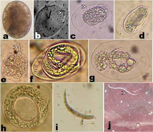
Initially the developing embryo grew up in size within the egg, and in the meanwhile hatching occurred. The second stage larvae attained larger size during migration, with development of a distinct cephalic tooth- like structure (Figure 5), within the tissues as these were gradually released from the cystic capsules that were extracted from the muscles of body of the infected fish. Thus these larvae migrated within the muscles of body, and finally entered venules and capillaries to find passage to the specially formed cavities with hepatic fluid within liver tissues of the same host. They rapidly grew in size to moult into third stage larvae. The advanced third stage larvae were then transformed into enormous-sized adults that attained maturity in liver and were lodged in a cavity within the tissues of liver, that had liquid in which the mature worms were drawned. The mature worms could be extracted from the cavity for collections and further study, by teasing the tissues of liver. The adult females laid eggs in the lumen of the cavity within tissues of liver, where hatching occurred in hepatic fluid within the cavity inside tissues of liver. The hatched larvae as well as eggs migrated through venules and capillaries of the hepatic- portal system of the infected fish to arrive at and get entangled into muscles tissues of practically every part of the body of infected fish. The eggs under various stages of development were also extracted from muscle tissues of fish during examination, while the moulting second stage larvae, early third stage larvae, and other developing stages were recovered during investigations by application of pepsin digestion method after FIBOZOPA Manual2 and modified Commission Direchne 84/319/EEC method.4

Figure 5 Life cycle stages [natural infections] (not to scale) of nematodes extracted by Pepsin Digestion Method. (a) egg, (b) polar plug on egg (c) egg with polar filament on one end (d) early development of egg showing cleavage (e) tooth in second stage larva, (f,g,h) moulting second stage larvae (i, j) early third stage larvae.
The description by original authors1 of Goezia bangladeshi in context with the larval stages of these nematodes have virtually been heavily laden with presumptions, based on facts that have really not been examined, but reported. This has led to unverified statements which have been discussed in the text. The description of such erroneous statements are quoted in Table 1. In light of these observations the taxonomic status of G. bangladeshi is circumspect unless appropriate description of the 2nd and 3rd stage larval forms of G bangladeshi are studied. Therefore, Goezia bangladeshi is termed as species inquirendae pending complete description of its larvae. The taxonomic position of the new worms accommodated under newly raised Tribe Indospineziinea, Subfamily Goeziinae, Family Raphidascarididae of the Order Raphidascaridoidea is thus justified and upheld.
|
The authors admitted that the worms studied and reported as “free larvae” were actually sexually immature juveniles, harboured by fish in the gut lumen. These were presumed to be the “larvae”, whereas no larvae were actually collected from the host. Some were also embedded in the intestinal wall. |
The morphology of adult worms of Indospinezia multispinatum separately from larvae from the original habitat, i.e. liver, were actually reported. The complete set of larval stages of nematodes were maintained and identified by application of Pepsin Digestion method. None Occurred in the intestine |
|
As a result Akther1 asserted that “The general biology and life cycle of G bangladeshi is likely to be similar to other Goezia species.”, though no actual life cycle stages were factually analysed. |
However, in the original form of I. multispinatum,3 the autogenicity in completion of the life cycle without the involvement of any intermedite host was actually recorded. |
|
On the contrary the larvae were presumed to have been ingested by a copepod, that might have developed to the third stage. |
No copepod intermediate hast was required in the life cycle of non- encapsulated form of I multispinatum3 the autogenicity where in developing larvae migrated through blood stream & returned to liver again. The larvae were neither encapsulated nor were embedded in gut wall, & instead were free inside muscular tissues of the same fish host in which they migrated from liver to other organs & progressed to transform into 3rd larval stage. |
|
The authors candidly admitted that no third stage larvae were found in T. ilisha. So the probability of moulting to produce worms beyond 3rd stage was again expressed, without evidence of occurrence of a boring tooth. |
The evidence of 2nd stage larva with boring tooth, supported with its LM picture has been presented. |
|
The intestinal caecum found poorly developed in Juveniles. |
A well developed intestinal caecum has been recorded in larvae examined. |
|
Authors themselves expressed opinion to conduct further studies to confirm their “probable” elements of observations. |
The evidence of larval tooth in 2nd stage larvae is presented with developed intestinal caecum & bifid ventricular appendix, that appeared later, the unique Anisakis features which were wanted in juveniles of G. bangladeshi. |
Table 1 Presumption based conclusions derived on larval stages in the life cycle of Goezia bangladeshi in the paper by Akther1 that led to erroneous interpretations and hence made the taxonomic position of Goezia bangladeshi circumspect unless appropriate larvae are studied
The authors are thankful to USIF, Aligarh Muslim University for SEM facility.
The authors declare that there are no conflicts of interest.

©2023 Yadav, et al. This is an open access article distributed under the terms of the, which permits unrestricted use, distribution, and build upon your work non-commercially.