International Journal of
eISSN: 2573-2838


Mini Review Volume 4 Issue 1
Department of Industrial & Information Engineering, University of Campania Luigi Vanvitelli, Italy
Correspondence: Luigi Zeni, University of Campania Luigi Vanvitelli, Department of Industrial & Information Engineering, Via Roma 29, 81031 Aversa, Italy, Tel +39 5010269
Received: December 20, 2017 | Published: January 11, 2018
Citation: Cennamo N, Zeni L. Bio and chemical sensing by a low cost plasmonic platform in plastic optical fibers. Int J Biosen Bioelectron. 2018;4(1):1–3. DOI: 10.15406/ijbsbe.2018.04.00086
A simple and low-cost approach to plasmonic sensing has been implemented exploiting a sensing multilayer deposited on a D-shaped Plastic Optical Fiber (POF) combined with a very easy experimental setup, based on a white light source and a spectrometer. This sensor system can be used for remote sensing, taking advantage of the capability of the optical fibers. When receptors are used for Bio/Chemicals detection, this sensing system can be exploited in different fields: medical diagnostics, environmental monitoring, industry, food safety and security. In the last five years, we implemented very important applications by specializing the same plasmonic POF platform with different kinds of receptors, such as Molecularly Imprinted Polymers (MIPs) and Bio-receptors. In the above applications, the experimental results showed that the set-up is very selective and able to sense very low concentrations.
Keywords: optical sensors, biosensors, chemical sensors, surface plasmon resonance, plastic optical fibers
POF, plastic optical fiber; MIP, molecularly imprinted polymer; SPR, surface plasmon resonance
Surface plasmon resonance (SPR) is widely used as a detection principle for many sensors operating in different application fields, such as bio and chemical sensing. When artificial receptors are used for bio/chemicals detection, the film on the metal surface (usually a gold surface) selectively recognizes and captures the analyte present in a liquid sample, so producing a local change in the refractive index at the metal surface. The value of the refractive index change depends on the structure of the analyte molecules.1 SPR biosensors based on Kretschmann and Otto configurations are usually bulky and require expensive optical equipment, it is not easy to miniaturize them and, in addition, their remote interrogation may be difficult to develop. Jorgenson et al. replaced the prism by a multimode optical fiber.2 The metal was deposited on the bare core of the fiber. The use of an optical fiber allows for remote sensing and may reduce the cost and the dimensions of the device. Due to the propagation of the light in the fiber, the angle of incidence on the metallic layer exceeds the critical angle, which depends on the refractive indices of both core and cladding components. Therefore SPR only exists for surrounding dielectrics whose refractive index lies in a narrow range. To overcome this drawback, Jorgenson et al. used a polychromatic light source and a spectrograph. This approach results in low cost, easy to implement device and can offer some attractive advantages such as the possibility to be used in the presence of flammable substances and hazardous environments, because of its electricity-free and remote sensing capabilities. Furthermore, because of the small size and non-invasive features, it can be used for medical (self-) diagnosis with the possibility to integrate SPR sensor platforms with optoelectronic devices, eventually leading to “lab on a chip”. In the scientific literature, many different configurations based on SPR in silica optical fibers, have been described.3-6 On the other hand, for SPR sensor platforms, POFs are especially advantageous due to their excellent flexibility, easy manipulation, great numerical aperture, large diameter, and the fact that plastic is able to withstand smaller bend radii than glass. Furthermore, the advantage of the POF sensors is that they are simpler to manufacture than those based on silica optical fibers.7
A versatile and low-cost optical sensor platform
Our optical platform is realized by removing the cladding of POF (along half circumference), spin coating a buffer layer on the exposed core and finally sputtering a thin gold film. The size of the POF is 980 μm of core (1 mm in diameter), whereas the multilayer on D-shaped POF presents a thickness of the buffer layer of about 1.5 μm and a thin gold film of 60 nm. The sensing region is about 10 mm in length. The buffer layer is made of photoresist (Microposit S1813), with a refractive index greater than that of the POF core. This buffer layer improves the performances of the SPR sensor and the adhesion of the gold film on the platform.7 In the visible range of interest, the refractive indices of the optical materials are about 1.49 for POF core (PMMA), 1.41 for cladding (fluorinated polymer) and 1.61 for buffer layer (Microposit S1813). Figure 1 shows the experimental setup for the SPR-POF sensor. It is composed by a halogen lamp, as the light source, the SPR-POF sensor and a spectrometer connected to a Laptop.
SPR-POF platform combined with receptors
Interesting results have been achieved in fields of medical diagnostics for cancer bio-markers detection8 and Celiac disease antigens monitoring,8 security with the detection of trinitrotoluene,9 environmental monitoring with the detection of perfluorinated compounds,10 food safety with the detection of Butanal,11 and industrial applications with the monitoring of furfural concentration in power transformers insulating oil.12
In this section we want to focus the attention on examples of bio-receptors, deposited on the SPR-POF platform, used in two medical diagnostic applications.
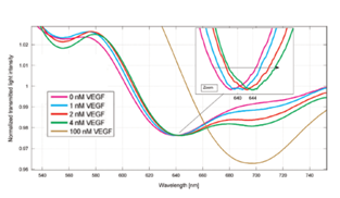
In the first case a DNA aptamer receptor, used for the detection of Vascular endothelial growth factor (VEGF), a circulating protein potentially associated with cancer, is reviewed.13 DNA aptamers are short oligonucleotide sequences that bind to non-nucleic acid targets with high affinity and specificity. The gold film of the SPR-POF sensor was firstly cleaned with an argon plasma, applying 6.8W of power to the RF coil for one minute to remove organic contaminants. Before the functionalization process, aptamers were subjected to thermal shock (95 °C for 1min, ice for 10min) in order to unfold the sequence strands and make the dithiol groups available for the immobilization reaction. The functionalization procedure was then accomplished by immersion into a 1 µM aptamer solution in 1 M potassium phosphate buffer pH 7 for one hour. To obtain Apt–MPET, a passivation step in 1mM mercaptoethanol solution in the same buffer for 30 min was performed after the functionalization with the aptamer. After washing in buffer and finally in ultrapure MilliQ water, the aptasensor was ready to use. In Figure 2 the transmission spectra of the SPR biosensor, obtained by contacting solutions at increasing concentrations of VEGF in buffer solution in the range 0-100 nM, are shown.13

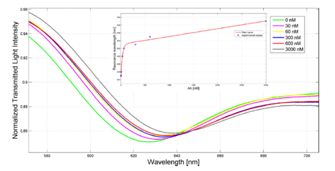
In the second case, a bio-receptor used for the diagnosis and/or follow-up of celiac disease, is reviewed.8 In particular, the POF-based sensor was used to monitor the formation of the transglutaminase/anti-transglutaminase antibodies, a new hallmark for the diagnosis of celiac disease. On the gold surface of the SPR-POF sensor, the guinea pig transglutaminase was immobilized by a simple approach. Before the functionalization, the gold surface of the chip was cleaned with ultrapure water and the deposition of droplets of a protein solution of 1 mg/ml (25µl) on the surface was performed. Then it was exposed to UV illumination (254 nm) for 10 min and incubated for 4 h at room temperature. When the surface was dried, a second deposition of protein was carried out (25µl of protein solution 1 mg/ml with UV illumination step of 10 min) and the chips were incubated over night at room temperature. The ability of the POF-sensor to detect the binding of anti-transglutaminase antibodies to the immobilized transglutaminase was studied.8 The obtained results indicated that the POF-SPR biosensor is able to sense the transglutaminase/antitransglutaminase complex in the range of concentrations between 30 nM and 3000 nM, as shown in Figure 3.

We presented a very simple, low-cost and versatile optical sensor platform, based on surface plasmon resonance (SPR) in a D–shaped POF. Several bio/chemical sensors, based on different kinds of receptors deposited on this SPR D-shaped POF platform, have already been implemented and new applications will be presented in the future. In particular the achieved results, relative to medical applications, indicate the ability to detect molecular complexes down to the nano-Molar range, exploiting different kind of bio-receptors and different functionalization procedures.
This work was partly supported by the Italian Ministry of Education, University and Research, by Regione Campania and by RSE – Ricerca Sistema Energetico - Italy.
The authors declare that they have no conflict of interest.

©2018 Cennamo, et al. This is an open access article distributed under the terms of the, which permits unrestricted use, distribution, and build upon your work non-commercially.