eISSN: 2471-0016


Case Report Volume 3 Issue 2
1Department of Laboratory Medicine1, NU Hospitals, India
1Department of Laboratory Medicine1, NU Hospitals, India
2Department of Urology, NU Hospitals, India
2Department of Urology, NU Hospitals, India
Correspondence: Kiran Krishne Gowda, Consultant Pathologist, NU Hospitals, Bangalore, India, Tel 9902313888
Received: January 01, 1971 | Published: November 11, 2016
Citation: Gowda KK, Matippa P. Parathyroid lipoadenoma: a diagnostic pitfall during frozen section evaluation of parathyroid lesions. Int Clin Pathol J. 2016;3(2):193–194. DOI: 10.15406/icpjl.2016.03.00070
Parathyroid lipoadenomas are a rare and benign variant of parathyroid adenomas that are defined morphologically by an abundance of adipocytes. The clinical manifestations and the laboratory findings are indistinguishable from those of the usual forms of parathyroid adenomas. Unlike parathyroid adenomas, parathyroid lipoadenomas can be functional or nonfunctional. We present a case of a functioning parathyroid lipoadenoma, which presented diagnostic difficulty on frozen section examination. The diagnosis was aided by intra-operative parathyroid hormone measurement.
Keywords: parathyroid; hyperparathyroidism, PTH, adenoma, parathyroid hormone, lipoadenoma, parathyroid lipoadenoma, PLA
Primary hyperparathyroidism is a disorder of inappropriate secretion of parathyroid hormone (PTH) from parathyroid tissue. Majority (80% to 85%) of primary hyperparathyroidism can be attributed to single enlarged parathyroid gland or adenoma.1 Parathyroid lipoadenoma (PLA) is extremely rare cause of hyperparathyroidism and differs from typical adenoma with regards to 1 major histologic criterion: this histologic variant is defined by presence of abundant stromal fat.2 Approximately 60 cases of parathyroid lipoadenoma have been documented in English literature.3-7 We present a rare case of hyper-functioning lipoadenoma, diagnosis of which was aided by intra-operative PTH measurement.
Case history
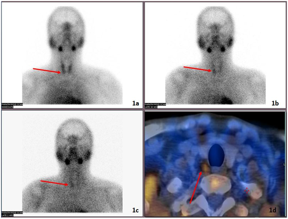
A 36 year old man with history of recurrent right ureteric calculi since 3 years presented with left ureteric colic. Computed Tomography scan of abdomen revealed calculus in left upper ureter measuring 7x4mm and hydronephrosis of left kidney. His biochemical tests revealed serum calcium of 10.01mg/dL (normal- 8.1 to 10.2mg/dL), and serum PTH of 257pg/mL (normal- 14 to 72pg/mL). In view of hyperparathyroidism, Sestamibi scan was done, which revealed solitary hyper functioning right inferior parathyroid gland in right trachea-esophageal groove (Figure 1).

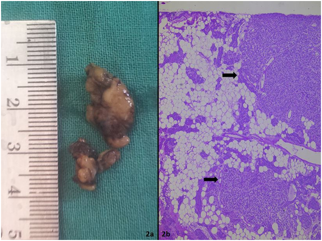
Suspecting an adenoma, right inferior parathyroid gland was excised along with left ureteroscopy and laser fragmentation of calculus. The parathyroid tissue sent to frozen section weighed 2000mg and measured 2.5x1.5x1cm (Figure 2a). Frozen section examination confirmed parathyroid tissue showing glandular elements admixed with adipocytes. Parathyroid tissue was labelled abnormal (lipoadenoma/lipohyperplasia) in view of weight and measurement. Rapid intraoperative PTH levels guided surgical procedure. Pre-incision PTH was 350.4pg/mL, while post-excision PTH was 18pg/mL (20 minutes after excision). A significant decrease of intraoperative PTH level of >50% from maximum baseline indicated successful excision of adenoma and negated need for further exploration.
A definitive diagnosis was established on histopathological examination of paraffin block of parathyroid tissue which revealed lipoadenoma with cords and nodular expansions of glandular elements (Figure 2) intimately admixed with adipocytes (60% of tumor). Minimal nuclear atypia was noted. However, no necrosis, fibrous band, mitosis, capsular or vascular invasion was noted. A peripheral rim of normal parathyroid tissue was noted.

Post-operatively patient had an uneventful recovery. At the time of last follow-up (6 months post surgery) his serum calcium was 8.9mg/dl (normal: 8.5-10.1mg/dl) and PTH was 41.4pg/ml (normal: 16-72pg/ml).
PLA is extremely rare, constituting <1% of all causes of hyperparathyroidism.3 It is a variant of adenoma characterized by abundance of adipocytes. Approximately 60cases of PLA have been documented in English literature.3–7 The current case would be 61st case. Similar to tumor in the current study more than 50% of PLA documented are functional.
Fat normally occupies approximately 25% of normal parathyroid gland and it may be increased by advanced age and obesity. The fat content of PLA is highly variable. Chow et al documented >50% fat in all 5 PLAs in their study.4 Seethala et al.3 reported a median fat content of 50% (range, 30%-70%) in 8 lipoadenomas. It was 60% in the current case. The origin of fatty tissue component remains unknown, but it has been speculated that same factors that drive the enlargement of parathyroid chief cells are responsible for enlargement of fatty component. The high fat content of lipoadenomas renders them difficult to localize using imaging techniques. Ultrasound has been reported to identify the tumors in 50% of cases, whereas sestamibi was successful in 71% in a series of 11cases.3 In our patient, sestamibi single photon emission computed tomography images showed increased activity in the right inferior parathyroid gland.
Lipoadenomas resemble normal parathyroid tissue because of their abundant stromal fat, which may cause diagnostic difficulty at the time of frozen section. Seethala et al evaluated the performance of frozen section in this setting and found that in all cases where frozen sections were performed, they were able to successfully identify that the parathyroid tissue removed was abnormal.3 They attributed their success largely to accurate measurement, weight, and good communication between the surgeon and pathologist as to how much of the gland was resected. This was the case in our patient. The parathyroid gland was labelled abnormal on frozen section, based on measurement, weight and complete excision.
Intraoperative PTH measurement is extremely useful in assessing the adequacy of excision of parathyroid adenoma. A significant decrease of the intraoperative PTH confirms that further exploration is not indicated.8 Fall in intraoperative PTH level of >50% from maximum baseline confirmed a parathyroid adenoma in the current study, despite the ambiguity in frozen section report. Final diagnosis was based on examination of paraffin block.
In summary, PLA are rare variants of parathyroid adenoma that pose difficulties on preoperative localization and pathologic evaluation especially frozen section. Awareness of this variant, adherence to ‘‘basics’’ of parathyroid gland assessment (weight, measurement, and good frozen section technique) along with intra-operative PTH measurement facilitate recognition during intraoperative setting.
None.
The author declares no conflict of interest.

©2016 Gowda, et al. This is an open access article distributed under the terms of the, which permits unrestricted use, distribution, and build upon your work non-commercially.