eISSN: 2373-6372


Case Report Volume 10 Issue 6
Department of Hepato-Gastro-Enterology, Mohammed VI University Hospital, Morocco
Correspondence: Lkousse Mohammed Amine, Department of Hepato-Gastro-Enterology, Mohammed VI University Hospital, University Mohammed VI, Oujda, Morocco, Tel +212611188323
Received: November 11, 2019 | Published: December 12, 2019
Citation: Lkousse MA, Gharbi K, farouki AEI, et al. Vulvar lesion in Crohn’s disease: a new case report. Gastroenterol Hepatol Open Access. 2019;10(6):306-307. DOI: 10.15406/ghoa.2019.10.00400
The dermatological manifestations are, with the osteo-articular, the most frequent of the extra intestinal attacks of Crohn disease (CD). While some of them evolve alongside the digestive disease, others evolve independently. Sometimes they can even precede the appearance of intestinal manifestations by several months, which then poses diagnostic problem.The recto-vaginal fistulas and ovarian involvement in CD have been widely reported in the literature.1,2 However, there are few series reporting the genital complications of CD, let alone regarding vulvar lesions.We report a new case of vulvar lesions in Crohn disease.
Keywords: crohn disease, recto-vaginal fistulas, vulvar lesions, granuloma, Anatomopathological study, Pelvic MRI
A 30-year-old divorced woman with 2 children was followed for CD evolving for 7years and treated with mesalazine. The patient did no longer consult for 5years. When she came back, the patient reported bloody diarrhea at the rate of 3 to 4 stools/day associated with tenesmus, evolving by flares/remission and pelvic pain.
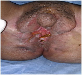
Clinically, the perineum was retracted with multiple external fistulous orifices, some of which are budding. There was also a remodeled vulva, a painful edema of the labia majora with a hypertrophied clitoris (Figure 1).Anatomopathological study of vulvar biopsies revealed a giganto-epithelioid granuloma without caseating necrosis, compatible with Crohn disease (Figure 2).

Figure 1Remodeled vulva, edema of the labia majora with hypertrophied clitoris and multiple perineal fistulas.
The colonoscopy showed a discontinuous inflammatory mucosa, with large ulcers up to left colic flexure. Above the mucosa was endoscopically normal. The anatomopathological examination of the colon biopsies gave the same results as those of the vulva in favor of Crohn disease.Pelvic MRI showed a large recto-vaginal fistula, multiple ano-perineal complex fistulas with perineal and pelvic infiltration, as well as circumferential and regular thickening of the extended rectum to the sigmoid colon (Figure 3).
Therapeutically, the patient received infliximab at 5mg/kg (including three injections at weeks 0, 2 and 6 and then every 8weeks) with onset of significant decrease in digestive, ano-perineal and vulvar manifestations. The surgical treatment had been proposed for the large recto-vaginal fistula.
The crohn disease is a chronic granulomatous inflammatory disease that can affect any part of the digestive tract. It is associated with frequent and polymorphic extra-intestinal manifestations, the most frequent of which are musculoskeletal, dermatological, and ophthalmological and hepatobiliary.The dermatological manifestations during CD include reactive dermatosis, autoimmune dermatosis, diseases related to nutritional deficiencies, dermatological lesions secondary to treatment and specific granulomatous lesions.3 The latter form can affect the genitals, and other parts of the skin.4,5 The vulvar CD was described for the first time by Park et al.6and there are few series in the literature reporting genital complications of CD, let alone vulvar lesions.The involvement of the vulva during CD can occur either by direct extension from the perineal region or by metastasis. Metastatic Crohn disease is defined as granulomatous lesions separated from the affected regions of the digestive tract by a territory of healthy skin.7,8
In a review of the literature, Andreani et al found that 91% of cases of vulvar CD were metastatic, while only 9% were contiguous from the perineal region.10In the same study, 25% of Vulvar CD cases had no digestive tract symptoms at the time of diagnosis.
Clinically, vulvar lesions can be associated to varying degrees with edema, papules, painful nodules, endovaginal fistulas, Bartholin's gland abscesses and ulcerations.1,2 The diagnosis must be evoked in the presence of deep vulvar linear ulcers called "knife-cut" or painful indurated labial edema, often asymmetrical.
The differential diagnoses of vulvar localization include sarcoidosis, tuberculosis, lymphogranulomavenereum, pyogenic infections, hidradenitissuppurativa, intertrigo and syphilitic lesions. The definitive diagnosis can only be made by performing a biopsy that reveals a non-caseating granulomatous lesion.9The natural evolution of vulvar CD is unpredictable. Although some cases resolve spontaneously, the majority of cases persist.12,13Due to its rarity, there are no randomized trials that suggest special treatment for vulvar Crohn disease.14The oral steroids were associated with long-term improvement,15 but other medications like oral metronidazole, azathioprine and 6-mercaptopurine also showed good results.16
The Infliximab has been effective in cases of vulvar CD resistant to other immunosuppressants, including prednisolone, azathioprine and oral cyclosporine.8.The adalimumab and certolizumab have also been shown to be effective in dermatological Crohn disease.17,18The surgery may be necessary for vulvar CD refractory to all medical treatments. Localized surgical excision frequently leads to localized recurrence and suboptimal wound healing. The radical vulvectomy is usually necessary in difficult cases.9
The vulvar localization is a rare extra-intestinal manifestation of Crohn disease. A multidisciplinary approach involving gastroenterologists, gynecologists, pathologists and dermatologists is recommended to identify this entity and to initiate appropriate treatment.As standardized therapy is lacking, the current therapeutic approach is case-by-case and can be escalated according to the severity of the disease.
None.
Authors declare that there is no conflict of interest.

©2019 Lkousse, et al. This is an open access article distributed under the terms of the, which permits unrestricted use, distribution, and build upon your work non-commercially.