eISSN: 2373-6372


Case Report Volume 14 Issue 3
1Department of, Surgery, Om Surgical Center & Maternity Home, India
2Department of Obstetrics and Gynaecolog, Om Surgical Center & Maternity Home, India
Correspondence: Pankaj Srivastava, Laparoscopic, Thoracic, Thoracoscopic & VATS Surgeon, Om Surgical Center & Maternity Home, SA 17/3, P-4, Sri Krishna Nagar, Paharia, Ghazipur Road,Varanasi, UP, INDIA. PIN-221007, Tel +91-542- 2586191; +919125679989
Received: June 06, 2023 | Published: June 30, 2023
Citation: Srivastava P, Srivastava S. Strawberry gallbladder: a velvety carpet of surgical anguish. Gastroenterol Hepatol Open Access. 2023;14(3):89-91. DOI: 10.15406/ghoa.2023.14.00552
Cholesterolosis is an uncommon pathologic finding of the gallbladder. The etiopathogenesis is not yet clear but it is proposed that it occurs due to increased cholesterol uptake from supersaturated bile. One must try to make a preoperative diagnosis by careful ultrasonography. Laparoscopic cholecystectomy is the gold standard treatment in symptomatic cases whereas asymptomatic patients can be treated conservatively as a case of functional dyspepsia.
Keywords: cholesterolosis, cholesterol stones, cholecystectomy, gallstones, strawberry, lipid crystals, cholesterol, cholecystitis
USG, ultrasonography; GB, gallbladder; CBD, common bile duct; CD, cystic duct; CA, cystic artery
Virchow first described Cholesterolosis in 1857 in his report on the role of the gallbladder in fat metabolism.1 Cholesterolosis is characterized by the accumulation of lipid-containing macrophages in the lamina propria of the gallbladder. It is a benign condition that is usually diagnosed incidentally during cholecystectomy or on ultrasonography (USG). Clinically, this may present as acalculous cholecystitis, calculous cholecystitis, and even pancreatitis.2
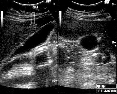
A 32-year-old male presented with mild to moderate pain in the right upper abdomen for 4 years that was increased after the spicy and fatty meal. He is a non-vegetarian, but mostly consumed vegetarian foods. During the past six months, he also experienced several episodes of vomiting after severe pain attacks. There was no history of any addiction like smoking, alcohol, etc. Gallbladder (GB) ultrasound showed well distended GB, normal wall thickness, and a single stone in its lumen (Figure 1). Acute calculous cholecystitis diagnosis was made based on history, examination and USG, and the patient was referred to us for laparoscopic cholecystectomy. When the patient was admitted, his vital signs were stable: his oral temperature was 98.40 F, pulse rate was 76 per minute, blood pressure was 110/70 mmHg, and his respiratory rate was 18 per minute. On examination, the right hypochondrium was tender. On deep palpation, the pain got aggravated and no lump or organomegaly was detected. The systemic examination was normal and no abnormality could be detected. Chest X-ray and ECG were also normal. The laboratory blood biochemistry and serology parameters were also within normal limits.
The patient was taken up for laparoscopic cholecystectomy under general anesthesia. The Patient was kept supine and standard four ports were introduced. The laparoscopic examination revealed a few flimsy adhesions around the gallbladder which were successfully taken off by the meticulous adhesiolysis. After proper dissection of the common bile duct (CBD), cystic duct (CD), and cystic artery (CA), CD and CA were clipped with LT300 titanium Ligaclips as standard procedure. The complete excision of the gallbladder was done. Peritoneal washing was done with normal saline and the GB bed was checked for hemostasis. 18 Fr abdominal drain was put near the GB fossa and port closure was done in layers. Extubation was successful after the operation and the patient was comfortable on room air. GB specimen was sent for histopathologic examination. No intraoperative or postoperative complication was noticed. The abdominal drain was taken out on next day and the patient was discharged. The patient was thoroughly followed up for two months.
The GB was 4.5cm long and 1.5cm wide on gross examination. The attached segment of the cystic duct was 0.2cm long and 0.3cm in diameter. The lymph node was absent. The serosa of the gallbladder is greyish-white, smooth, and shiny (Figure 2). The hepatic resection surface is tan-brown, with associated cautery artifacts. Single stone present in the lumen. The gallbladder wall measures 0.3cm in thickness and is grossly unremarkable. The mucosa is tan-brown and velvety, without polyps or other lesions.
Histologic examination showed a gallbladder wall with moderate chronic inflammatory infiltrate and foamy macrophages in lamina propria. There was thickening of the muscularis. There were no features suggestive of malignancy in the processed section. Rokitansky Aschoff sinuses are seen (Figure 3). The final diagnosis of the strawberry gallbladder was made.
Cholesterolosis is an incidental finding usually after the cholecystectomy when the GB is cut to see the inner surface, stones and, tumors if any. The exact pathogenesis is still not known but it is supposed to be related to the cholesterol metabolism. Macroscopically lipid deposits appear as yellow flecks against dark yellow to green background, thus earning the nickname ‘Strawberry Gallbladder’.3
Accumulation of larger lipid aggregates can lead to polypoid growths that protrude into the lumen and are commonly known as cholesterol polyps, whereas these should be termed cholesterolosis polyps. Cholesterol esters and triglycerides when get accumulated can lead to increased lipid synthesis in the liver and increased absorption and esterification by the gallbladder. A normal gallbladder absorbs free and nonesterified cholesterol from bile. Macrophages phagocytosed the lipid droplets which are formed by the esterification of the cholesterol. Hence, the cholesterolosis is caused by increased cholesterol uptake from supersaturated bile.
Another school of thought is that cholesterolosis is caused by defects in macrophages that are no longer able to metabolize and excrete cholesterol absorbed from bile. Though the diagnosis by ultrasonography is difficult when these deposits get bigger in size, they can be well detected by the USG (Figure 4).4 The Spectrum of Clinical presentation is wide from the silent finding without any symptom to free-floating biliary sludge to pain in the right upper abdomen due to acalculous cholecystitis to calculous cholecystitis with cholesterol stones.5 Laparoscopic cholecystectomy should be considered as the treatment of choice in symptomatic patients of gallbladder cholesterolosis.

Figure 4 USG of Acalculus Cholecystitis with thickened GB wall studded with the cholesterol crystals..
Cholecystectomy is especially suitable for patients with biliary colic or pancreatitis because many patients with cholesterolosis improve after cholecystectomy.6,7 On the other hand, patients with non-specific dyspeptic symptoms but without symptoms of biliary colic should be managed conservatively since the pathogenesis of these symptoms is unclear and cholecystectomy may not relieve the symptoms. Such patients should be treated symptomatically, as are other patients with chronic functional dyspepsia.5
Though cholesterolosis is a rare benign condition of the gallbladder with uncertain etiopathogenesis, one should take of treating the condition by the gold standard “Laparoscopic Cholecystectomy”. The surgeons should try to clear the diagnosis by the USG especially in the acalculous conditions because these subset of patients can be well treated by the conservative management.
None.
We declare there are no conflicts of interest.
None.

©2023 Srivastava, et al. This is an open access article distributed under the terms of the, which permits unrestricted use, distribution, and build upon your work non-commercially.