eISSN: 2373-6372


Case Report Volume 12 Issue 3
1Department of Colorectal Surgery, University Hospital of Brasilia, Brazil
2School of Medicine, University of Brasilia, Brazil
3Department of Surgery (Coloproctology), University of Brasilia, Brazil
Correspondence:
Received: June 03, 2021 | Published: June 14, 2021
Citation: Martins BAA, Ferreira IAR, Oliveira PG, et al. Splenic infarction after colonoscopy: direct complication or unhappy coincidence? Gastroenterol Hepatol Open Access. 2021;12(3):98-99. DOI: 10.15406/ghoa.2021.12.00464
Introduction: Splenic infarction is a condition that occurs when the splenic artery or one of its branches become occluded and it may be rarely caused by abdominal procedures. We report the case of a 66-year-old male patient who presented splenic infarction after screening colonoscopy.
Case report: A 66-year-old male patient presented to the emergency department with a 6-day history of acute onset of the abdominal pain. The complaint initiated after screening colonoscopy. CT scan revealed signs of splenic infarction. Patient presented uneventful recovery with supportive care. Subsequent investigations revealed the diagnosis of atrial fibrillation.
Discussion: Splenic infarction typically manifests as abdominal pain in the upper left quadrant. Atrial fibrillation seems to be the main cause of embolic events that result in infarction of the spleen. In the present report, symptoms started after a colonoscopy what raises the possibility of a causal link between the endoscopic procedure and the splenic infarction.
Keywords: splenic infarction, colonoscopy, endoscopy, heart disease, blood pressure, antibiotics, analgesics, antiemetics
Splenic infarction is a condition that occurs when the splenic artery or one of its branches become occluded. It may be rarely caused by abdominal procedures such as gastrectomy, Nissen fundoplication, peritoneal dialysis and endovascular procedures.1 Although colonoscopy is a known cause of splenic rupture, cases of splenic infarction associated with this examination method have never been reported.2 Splenic infarction treatment ranges from expectant management with analgesia and hydration to splenectomy.3
In view of the relevance of this topic for professionals who perform endoscopic exams, we report a patient who presented with splenic infarction after screening colonoscopy.
A 66-year-old male presented to the emergency room with a 6-day history of moderate pain and tenderness in the upper left quadrant of the abdomen. Abdominal pain was initiated after screening and an uneventful colonoscopy, in which a 3mm sessile polyp was identified in the sigmoid colon. Cold forceps polypectomy was performed, and histopathological examination revealed tubular adenoma with low-grade dysplasia.
The patient is a former smoker, has had chronic Chagas’ heart disease for 34years and has no history of abdominal surgery. He uses captopril, propranolol, amlodipine and hydrochlorothiazide daily.
Physical examination showed a heart rate of 60 beats/min, blood pressure of 150/92mmHg, respiratory rate of 18 breaths/min, and oxygen saturation of 96% on room air. The abdominal examination was significant for mild pain in the left abdomen, and there were no signs of peritoneal irritation. Laboratory tests showed a haemoglobin level of 16.7mg/dL, a white blood cell count of 9.0×10⁹/L and a C-reactive protein level of 9.18mg/dL.
Once admitted to the emergency care unit, an acute abdominal series and a contrast-enhanced abdominal CT were requested. The acute abdominal series revealed only a small left-sided pleural effusion. Abdominal CT was not performed at the time because the hospital scanner was under maintenance. The patient was admitted to the ward, and supportive care was started with the administration of antibiotics, analgesics and antiemetics. With complete resolution of pain intensity and an improved general condition, the patient was discharged one day after admission.
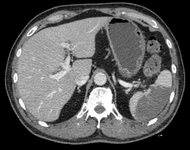
Six days after patient discharge, contrast-enhanced abdominal CT was performed and revealed a normal volume spleen with a large and peripheral wedge-shaped hypoenhancing area compatible with splenic infarction (Figure 1).

Figure 1 Abdominal CT scan demonstrating a large peripheral wedge-shaped hypoenhancing region in the spleen.
In a consultation held three months after his admission, the patient had no complaints and reported good recovery. He was referred to other specialists for investigations of silent diseases that could be related to the episode and an electrocardiogram revealed the diagnosis of atrial fibrillation.
Splenic infarction typically manifests as abdominal pain in the upper left quadrant. Other less common symptoms and signs are fever, chills, nausea, vomiting and splenomegaly. Laboratorial findings are elevated lactate dehydrogenase, alkaline phosphatase and leukocytosis.1,4,5 Abdominal CT scan is imperative in the evaluation of suspected splenic infarction and classically reveals as a peripheral wedge-shaped area with the broad base towards the splenic capsule.6,7
The incidence of this event is unknown. Schattner et al. found that splenic infarction represented only 0,016% of admissions over 10years in a single academic hospital.1 It can affect individuals of all ages. Its aetiology is generally linked to haematological disorders when it affects patients younger than 40years old, while in patients older than 40years old, embolic events are more prevalent.8 Atrial fibrillation followed by infectious endocarditis and atherosclerotic lesions in the aorta appear to be important causes of embolic events that cause infarction of the spleen.1,4,9
In the present case, the symptoms started after the colonoscopy, which raised the possibility of a causal link between the endoscopic procedure and the splenic event. However, it is extremely important to consider the patient’s history to raise the most likely diagnostic hypothesis. The patient has Chagas’ heart disease, a disease known to be associated with atrial fibrillation, as well as other heart diseases.10,11 Atrial fibrillation, which had not yet been diagnosed in this patient at the time, is the main cause of splenic infarction and increases the risk of embolic episodes involving several organs. A hypothesis that may justify the triggering of splenic infarction by colonoscopy would be the mobilization of emboli of the splenic nutritional vessels or in situ thrombosis by the traction caused by the passage of the colonoscope through the splenic flexure.
The initial treatment for splenic infarction includes clinical support, intravenous hydration and analgesia. Anticoagulation is used in cases secondary to thromboembolism, and splenectomy should be considered in patients with persistent symptoms.3,4,9 With a benign course, most patients recover well if properly treated. Even so, splenic infarction can be a diagnostic clue for the discovery of underlying conditions such as atrial fibrillation, polycythaemia vera or antiphospholipid syndrome. Some of the rare complications of splenic infarction are subcapsular or peritoneal haemorrhage and the formation of pseudocysts and abscesses.1,4,9 In the presented case, patient showed good recovery after conservative support, including intravenous rehydration, analgesia, antiemetics and antibiotics.
Colonoscopy is a widely used test for the diagnosis and treatment of multiple conditions in the colon, and its increasing demand increases the frequency of complications. Among its best-known complications are bleeding and colon perforation.12
Splenic laceration is a rare and under-detected complication of colonoscopy. This traumatic injury can be associated with intubation difficulty, looping of the instrument, adhesions between the colon and spleen, traction on the splenocolic ligament and the presence of polyps or masses at the splenic flexure.13–15 Despite several reports of splenic laceration after colonoscopy in the literature, we did not find any description of association between colonoscopy and splenic infarction. This case report raises the possibility of a causal link between the performance of colonoscopies and the occurrence of splenic infarction.
None.
The authors declare no conflicts of interest related to this article.
None.

©2021 Martins, et al. This is an open access article distributed under the terms of the, which permits unrestricted use, distribution, and build upon your work non-commercially.