eISSN: 2373-6372


Case Report Volume 14 Issue 1
Department of Infectiology, Aboubekr Belkaid University, Algeria
Correspondence: Badla Y, Faculty of Medicine, Department of Infectiology, Aboubekr Belkaid University, Algeria
Received: January 04, 2023 | Published: February 13, 2023
Citation: Badla Y, Benzeroual A, Rebib N. Relapsed cholangitis revealing hepatic distomatosis in western Algeria apropos of a case. Gastroenterol Hepatol Open Access. 2023;14(1):19-21. DOI: 10.15406/ghoa.2023.14.00537
Fascioliasis is a worldwide but unevenly distributed zoonosis caused by the trematode Fasciola hepatica that infects domesticated herbivores. Fascioliasis also occurs accidentally in humans through ingestion of freshwater or aquatic plants laden with metacercariae. Human infections are common in developing countries and not uncommon in Europe, and rare in Algeria. The clinical evolution has been classically described in two phases: an acute phase of hepatic parenchymal invasion of an immature worm larva (parenchymal phase) and a stationary phase after stay in the bile duct and egg production (ductal phase). We report a case of a 50-year-old man from Tlemcen, western Algeria, with cholangitis (liver disorder, abdominal pain and jaundice). The diagnosis was confirmed by serology. The serological examination (Western Blot) was positive and the results of magnetic resonance imaging were compatible with fascioliasis.
Keywords: cholangitis, fasciola hepatica, watercress, jaundice
Distomatoses are cosmopolitan zoonoses caused by trematodes:Fasciola hepatica and F gigantica.1 Global prevalence has exceeded three million cases.2
In Algeria, only hepatobiliary distomatosis, or Fasciola hepatica fasciolosis, called large liver fluke, is pathogenic for humans. These are rare and sporadic cases.
The disease presents in the invasion phase with non-specific digestive disorders, asthenia, myalgia. Complications are mechanical and inflammatory: retentional jaundice, hepatic colic attacks, access of cholangitis, cholecystitis.
The biological diagnosis is essentially based on the search for antibodies on serum. Diagnosis of a suspicion of distomatosis requires the search for circulating antibodies by immunoenzymatic technique (EIA or "ELISA") or indirect hemagglutination (HAI) and by immunoblotting (IE "Western blot").3 The treatment of fascioliasis is based on 2 doses of 10mg/kg of triclabendazole administered 12hours apart, orally. Nitazoxanide 500mg orally twice a day for 7 days may be effective, but dataare limited. We are going to report a case of hepatic distomatosis due to Fascioloa hepatica diagnosed following cholangitis
A 50-year-old man, married father of 03 children, originally from Beni Senouss in Tlemcen, director of a professional company, with no known medical and surgical history consulted within the infectious disease department in July 2021 for: fever, sweats, dry cough, abdominal pain and progressive constipation for 10days
Clinical examination is normal
Radiologically: Chest X-ray and abdominal ultrasound are normal
Biologically: normal hemogram, C-reactive protein CRP at 342mg/L, sedimentation rate at 68 at the first hour
Serology by immune blot (Western Blot) confirmed the diagnosis of hepatic distomatosis due to fasciola hepatica (Figure 1).

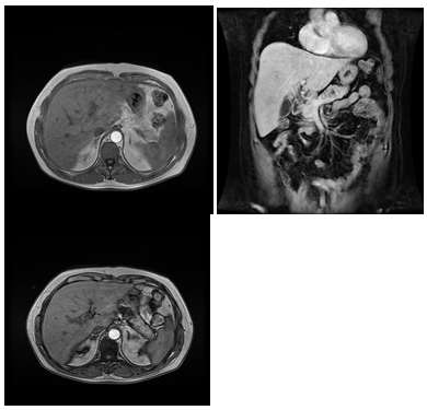
Figure 2 Hepatic magnetic resonance imaging (MRI) which shows harmonious hepatomegaly with hypovascular areas and vermicular aspects.
The patient was treated with triclabendazole 10mg/kg for three days with a good clinical and biological evolution. Three months later, the patient repeated the same clinical and biological picture with fever, sweating, hepatic colic, weight loss of five kilograms, hypereosinophilia at 6500 elements per milliliter, hepatic cytolysis three times normal and CRP at 196mg/l. The Hepatic magnetic resonance imaging (MRI) showed an heterogeneous harmonious hepatomegaly by the presence of a poorly circumscribed hypovascular area most likely corresponding to a vermicular appearance with signs of cholangitis. Stool parasitology revealed viable eggs of Fasciola hepatica, the patient was treated a second time by Praziquantel in trios taken for a single day with antihistamines.
There were no adverse effects.
Hepatobiliary distomatosis or fasciolosis is a parasitic condition due to the invasion of the liver and bile ducts by a species of Trematode, Fasciolahepatica commonly called Great liver fluke, it is a zoonosis which mainly affects ruminants. Humans become infected by ingesting plants contaminated with metacercariae.4 which is very popular in all regions of herbivore breeding,5 with the exception of cold areas such as Canada, northern Scandinavia, Iceland and Siberia.6 Foodborne trematodiasis is an emerging public health problem, particularly in Southeast Asia and the Western Pacific region.7 There are two main species affecting cattle in Algeria, namely Fasciola hepatica which causes a trematode disease that is endemic in our country and Dicrocoelium dendriticum or small liver fluke.6
In Algeria, studies on hepatobiliary distomatosis caused by Fasciola hepatica and its vector, although they date back to the 1800s, remain insufficient compared to those carried out, for example, in Europe. Cases of human distomatosis have been reported by
SENEVET and CHAMPAGNE in 1928 and 1929 (8) and by Guy et al. in 1969 5.9 The territory of Beni Snous is located in the mountains of Tlemcen, 41km southwest of Tlemcen, has a hot Mediterranean climate with dry summer (Figure 03).
The clinical picture of our patient was more or less typical with the association of infectious, respiratory and hepatic syndrome, we were influenced by the occurrence during the third wave of COVID 19 which caused a delay in diagnosis.
The contamination was summer unlike the cases published by D. Szymkowisk (5) where the contamination was in winter.
Clinically, our case was comparable to the two cases published by Seyed Farshi10 in IRAN who were operated for suspected cholelithiasis.
As soon as the diagnosis of hepatic distomatosis was suspected, we proceeded to an investigation in the entourage of our patient. We know, in fact, the familial character of the disease, due to the fact that it is at the family table that the infestation occurs. The disease can also present a frankly epidemic character. It spreads all the more as infesting watercress becomes more popular and more successful within the population considered. Our patient had stayed around a reviere in beni senouss and had consumed watercress with other family members composed of 07 members the cases of hepatic distomatosis thus detected were trivial in terms of their clinical and biological expression, and in terms of their evolution, also trivial, towards recovery, under the effect of treatment (Albendazol)
Two brothers 47 years old and 43 years old who had the same symptomatology but without relapses
The great polymorphism of the clinical picture presented by the parasitized people around this small epidemic has been reported by several series of literatures such as that of Rondelaud.11
Human distomatosis is rare in Algeria12 like the Maghreb countries and in France, but frequent in certain countries such as Egypt13 where a seroprevalence of human hepatic distomatosis has reached 18%.14 However, several studies have revealed a considerable frequency of animal hepatic distomatosis15 we can wonder about the causes behind this decrease in human cases, especially when we know that the animal reservoir of the disease is important (cattle) the change in diet could constitute a valid hypothesis to explain thereby.
None.
The authors declare that they have no known competing financial interests or personal relationships which might appear to influence this article.
None.

©2023 Badla, et al. This is an open access article distributed under the terms of the, which permits unrestricted use, distribution, and build upon your work non-commercially.