eISSN: 2373-6372


Case Report Volume 4 Issue 5
Department of Pediatric gastroenterology, hepatology and nutrition, New York Medical College, USA
Correspondence: Stuart H Berezin, Department of Pediatric gastroenterology, hepatology and nutrition, Maria Fareri Children?s Hospital, Westchester Medical Center, New York Medical College, Suit 201. 503 Grasslands road, Valhalla, New York, USA
Received: November 17, 2015 | Published: April 25, 2016
Citation: Pandey A, Berezin SH, Bamji N, Debelenko L (2016) Rectal Carcinoid Tumor in Adolescent Boy. Gastroenterol Hepatol Open Access 4(5): 00113. DOI: 10.15406/ghoa.2016.04.00113
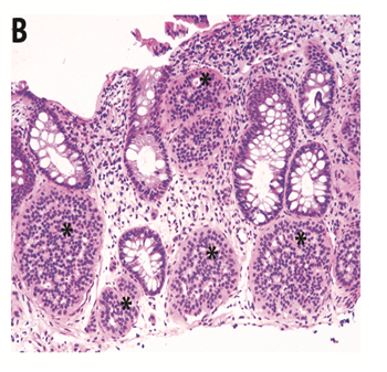
The patient is a 17 year old male, who was diagnosed with ulcerative colitis 10 years ago. His treatment was balsalazide. A surveillance colonoscopy was normal except for a small 0.2x 0.2 cms nodular lesion noticed at 15 cms from the anal verge Figure 1(A&B). The nodular lesion was removed by cold biopsy forceps. Biopsies of the terminal ileum were normal. Colonic histology showed chronic minimally active colitis. Histology of the nodular lesion confirmed the diagnosis of rectal carcinoid tumor. Magnetic resonance (MR) enterography and ultrasonography of liver and pancreas were normal. The patient subsequently had an endoscopic ultrasonography with Doppler which did not show any submucosal lesion or rectal wall thickening. There was no indication of residual carcinoid tumor.

Figure 1A Gross appearance of rectal carcinoid tumor: Colonoscopic appearance of carcinoid showing nodular lesion in rectum.

Figure 1B Histology of rectal mucosa showing scattered multiple nests of uniform round cells arranged in lace-like pseudorosetting pattern in lamina propria. H&E; original magnification X100.
Neuroendocrine tumors are rare. The incidence is 2.8 cases per million under the age of 30 years. Carcinoid is the most common neuroendocrine tumor of the GI tract.1 Tumor size larger than 10 mm, presence of central depression, increasing depth of tumor invasion, lymphatic and venous invasion were significantly associated with a higher incidence of lymph node metastasis.2 Five year survival rate has been reported to range from 54% to 73% for lymph node positive rectal carcinoid disease even without distant metastases.3 Most patients are asymptomatic but rectal carcinoids can present with rectal bleeding, abdominal pain, diarrhea and flushing of the skin. Rectal carcinoids are essentially chemo resistant.4 Surgical resection of metastatic lesions improves survival.5 Preoperative assessment with EUS for rectal carcinoids is effective to determine depth of invasion.6
None.
The author declares there is no conflict of interest.
None.

©2016 Pandey, et al. This is an open access article distributed under the terms of the, which permits unrestricted use, distribution, and build upon your work non-commercially.