eISSN: 2373-6372


Review Article Volume 13 Issue 2
Department of General Surgery, Hospital Provincial Docente Clínico Quirúrgico Manuel Ascunce Domenech, Cuba
Correspondence: Héctor Alejandro Céspedes Rodríguez, Department of General Surgery, Hospital Provincial Docente Clínico Quirúrgico Manuel Ascunce Domenech, Cuba, Tel 58360607
Received: February 08, 2022 | Published: March 28, 2022
Citation: Rodríguez HAC, Pérez RF. Malignant duodenocolic fistula as a form of presentation of right colon cancer. Case report. Gastroenterol Hepatol Open Access. 2022;13(2):60-61. DOI: 10.15406/ghoa.2022.13.00495
Malignant duodenocolic fistula in an unusual presentation internal fistula, whose main symptoms are abdominal pain, weight loss, halitosis, fecaloid vomiting and diarrhea. We report the case of a 79-year-old patient with diarrhea, fecaloid vomiting and significant weight loss, who is surgically intervened with the diagnosis of duodenocolic fistula, performing right hemicolectomy, partial duodenectomy, transverse duodenorphy as definitive treatment.
Keywords: duodenocolic, malignant fistula, fecaloid vomiting
Duodenocolic fistula is a rare presentation of colorectal cancer. The first report of a duodenocolic fistula of malignant cause was made by Haldane in 1862 published Edinburgh Medical Journal.1 This occurs when a neoplasm of the colon is locally advanced, with a communication occurring between the second portion of the duodenum and the hepatic flexure of the colon, establishing an internal fistula, which causes serious metabolic and nutritional alterations due to the bypass. The main symptoms and signs are abdominal pain, diarrhea, fecaloid or fecal vomiting, halitosis, and weight loss.2 Given the low prevalence of this pathology and little experience in our center, we report a patient with a duodenocolic fistula due to colorectal cancer.
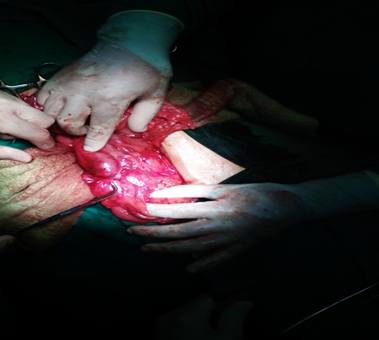
79-year-old, white, male patients with a history of high blood pressure. Who is admitted to the internal medicine service of the Manuel Ascunce Domenech hospital, due to presenting diarrhea, halitosis and weight loss during his stay in the ward, has fecaloid vomiting for which he is evaluated by surgery and before the absence of occlusive syndrome, we indicate endoscopy superior which reports at the level of the second duodenal portion abnormal communication with the colon is observed and we completed the study with esophagus-stomach-duodenum and contrasted CT, both observing how the right colon was filled early. It is concluded as a duodenocolic fistula (Figure 1 & 2), performing with this diagnosis (Figure 3) right hemicolectomy, partial duodenectomy, closure of the defect in a transverse way after the Horsley method and we performed choledocostomy by T-tube and jejunostomy was used for the Decompressions the first 72hours then for food purposes at 10days he was discharged after checking the suture with water-soluble contrast.

Figure 1 This image shows the tumor in the transverse colon approximately 12cm from the hepatic flexure, which infiltrates an area between the 2nd and 3rd portions of the duodenum.
Duodenocolic fistula usually presents between the sixth and seventh decade of life without distinction of sex or race. The incidence of duodenocolic fistula in the United States is 1 in 900 patients with colorectal cancer. Hershenson3 documented a case after performing 8100 autopsies on patients who died from colon cancer. These are classified into two primary groups that correspond to postoperative and secondary to the complication of an inflammatory process, this group in turn is subdivided into benign and malignant, the latter is less common than benign, the first report of a benign coloduodenal fistula is performed by Sanderson in 1863 caused by the rupture of a duodenal diverticulum.4
Among the causes of benign duodenocolic fistula include duodenal ulcer, Crohn's disease, migration Gallstones, diverticular disease of the colon and duodenal, rupture of a pancreatic pseudocyst, the most unusual cause reported is biliary stent migration in a patient with pancreatic cancer.5 As malignant causes we have colon and duodenal cancer and some authors mention gallbladder and esophageal cancer.6
Patients with a duodenocolic fistula will present symptoms and signs that will be dependent on the primary lesion, typical of the fistula and the presence of metastatic disease. Dependent on the fistula are diarrhea, fecaloid or truly fecal vomiting, halitosis, and significant weight loss. Diarrhea is the result of the presence of colonic bacteria in the upper tract which is sterile, the irritative effect of bile salts in the colon and the short intestine mechanism due to said communication. Depending on the primary lesion, we have abdominal pain, a palpable tumor in the right upper quadrant and anemia, the latter two are only present in 10% of patients with a malignant entero-enteric fistula.6-9 The diagnosis of duodenocolic fistula can be established with an esophagus-stomach -duodenum, upper endoscopy and a contrasted abdominal computed tomography which, in addition to defining the location, will establish the presence of metastasis.7
Treatment of malignant duodenocolic fistula depends on the extent of the primary tumor, the presence of metastases, and the general conditions of the patient. There are three standard procedures for the treatment of this fistula, two for curative purposes, which are right hemicolectomy with cephalic pancreatoduodenectomy or partial duodenectomy with closure of the defect7–9 and we have a gastrojejunostomy with ileotransversostomy for palliative purposes. For many authors, right hemicolectomy and partial duodenectomy is considered the treatment of choice due to the low mortality compared with the whipple procedure.9 Izumi et al.8 in a study of 34 cases of malignant duodenocolic fistula in Japan, performed the Whipple procedure plus right hemicolectomy as an alternative treatment for this entity, with a survival of 7days to 4years. Chang et al.9 treated 20 cases with right hemicolectomy and partial duodenectomy. Ellis et al.10 used jejunal serosa for the closure of the duodenal defect. We consider that in these cases where there is a duodenocolic fistula, whether of malignant or benign origin, where surgery such as the Whipple procedure is not performed, it is convenient to carry out two procedures, one to perform a jejunostomy for feeding and a prior choledocostomy by means of a T-tube. choledochotomy to adequately identify the papilla and avoid inverted injury and to decompress the bile duct before a primary duodenal suture, which would help improve the patient's evolution.
Malignant duodenocolic fistula is a rare entity that occurs in carcinoma of the hepatic flexure of the colon in its advanced form, generally between the sixth and seventh decade of life. The standard treatment is right hemicolectomy with partial duodenectomy and closure of the duodenal defect in a transverse manner.
None.
None.

©2022 Rodríguez, et al. This is an open access article distributed under the terms of the, which permits unrestricted use, distribution, and build upon your work non-commercially.