eISSN: 2373-6372


Case Report Volume 12 Issue 6
Department Gastroenterology, Faculdade de Medicina do ABC, Brazil
Correspondence: Wilson R Catapani, Faculdade de Medicina do ABC - Gastroenterology Av. Lauro Gomes 2000/ Santo André/SP 09060-650, Brazil
Received: November 20, 2021 | Published: December 16, 2021
Citation: Dassoler R, Catapani WR. Life-threatening, acute lower gastrointestinal hemorrhage as the first presentation of Crohn’s disease. Gastroenterol Hepatol Open Access. 2021;12(6):196-197. DOI: 10.15406/ghoa.2021.12.00484
Most patients with Crohn's disease usually present with mild gastrointestinal bleeding, and severe GI hemorrhage is rare. We report a case of a young male who presented with life-threatening lower gastrointestinal hemorrhage as the first manifestation of Crohn's disease.
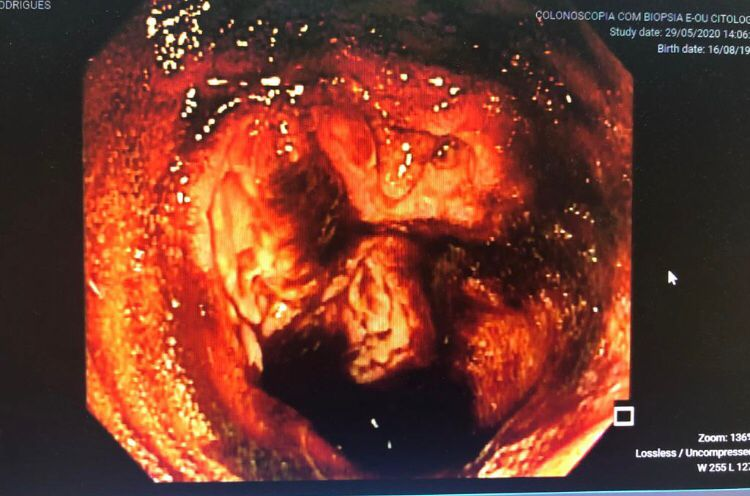
The patient gave informed consent for this report. He is a 24 years old computer technician with no previous comorbidities. The patient started with mild and intermittent diffuse abdominal pain in May 2020, evolving to continuous and severe pain, prompting a medical consultation. He underwent a computed tomography scan of the abdomen, which showed parietal thickening and hyperenhancement in the distal ileum, with engorgement of vasa recta in the mesentery, suggesting an inflammatory/infectious process. The patient was admitted to the hospital, receiving treatment with ciprofloxacin and metronidazole, initially diagnosed with infectious diarrhea. Upper digestive endoscopy was performed, which resulted in normal. However, after three days, he started with bloody diarrhea and worsening abdominal pain. A colonoscopy was performed, disclosing a friable mucosa with cobblestones, deep ulcers covered by fibrin intermingled with normal mucosa, and large amounts of blood in the colon and ileum (Figure 1).


Figure 1 Colonoscopy showing a friable mucosa with cobblestones, deep ulcers covered by fibrin and large amounts of blood in the colon and ileum.
The patient evolved with massive enterorrhagia, weakness, syncope, and severe anemia requiring transfer to the intensive care unit. At the ICU, the condition progressed to hemorrhagic shock, hemodynamic instability, and sensorium depression, requiring intubation, mechanical ventilation, and blood transfusion. Arteriography showed active digestive hemorrhage in the ileal branch of the mesenteric artery, which was embolized. After the procedure, the patient was extubated but maintained vasoactive drugs and a hemoglobin of 8.1g/dL. Subsequently, the next day he presented with a voluminous enterorrhagia and a drop in hemoglobin to 6.0g/dL. New arteriography was performed that showed active arterial hemorrhage, a new embolization was done, with cessation of bleeding. After 12hours of this procedure, the patient developed new episodes of enterorrhagia and a drop in hemoglobin to 4.4.g%. At this moment, we opted for an indication of surgery (enterectomy), with the removal of 60 cm of the ileum and end-to-side anastomosis. An anatomopathological report of the surgical specimen showed chronic transmural ileitis with architectural distortion of villi and crypts, follicular lymphoid hyperplasia with lymphoid follicles in the submucosa, and muscularis propria, in addition to deep non-caseous epithelioid granulomas in muscular propria and submucosa. Staining for fungus and tuberculosis by Grocott and Ziehl Neelsen were negative. The patient was discharged from the hospital and two months later had new episodes of enterorrhagia, treated with a colonoscopy and placing hemoclips in the region of the anastomosis.
In the first ambulatory consultation after discharge from the hospital, he started treatment with infliximab 5mg/kg. In his last ambulatory consultation (September 2021), the patient was asymptomatic and had a control colonoscopy showing no lesions and a healed mucosa, maintaining infliximab 5mg/kg every eight weeks.
Severe gastrointestinal bleeding in Crohn's Disease is a rare complication, with an incidence of 0.6 to 4%, being described most often in the literature through case reports. It is a diagnostic and therapeutic challenge due to the reduced number of cases, short follow-up time, difficulty in accurately identifying the source of bleeding, and the presence of other complications such as stenosis that can compromise the endoscopic evaluation, an increased risk of recurrent bleeding and, finally, limitations in the clinical data, making it difficult to reach a consensus on the definitive diagnosis and treatment modality of this condition.
Early reports (1996) pointed out that, among five hundred and one patients with Crohn's disease, studied during 20 years, ten (1,9%) had bleeding significant enough to require a blood transfusion. In most patients (90%), the source of bleeding was colonic.1
In another series from the USA, four patients of 631 admissions (0.6 percent) presented with life-threatening gastrointestinal hemorrhage. To these four cases, the authors added another thirty-four similar cases from the medical literature. The results showed a preponderance of young men with an average age of 35years, with the majority having Crohn's disease for an average of 4.6years. The five cases of exsanguination (13%) were all men with known Crohn's disease involving the ileum alone or in part. Mesenteric arteriography was positive in 17 patients, providing precise preoperative localization resulting in no mortality in this group.2
In hemorrhagic Crohn's disease, isolated colonic bleeding is the most common, between 3% to 50%, diffuse ileocolonic lesions between 22.7% to 68.5%, and other smaller parts of the intestine 19% to 66%, with jejunal and superior bleeding being infrequent. Our case report shows ileal bleeding that is difficult to control utilizing embolization, evolving to surgical treatment. In addition, bleeding is seen on average five years after diagnosing Crohn's disease, different from what is presented in this report.
Kim et al. studied risk factors for acute lower gastrointestinal bleeding in Crohn's disease. They found a cumulative probability of bleeding after diagnosis being 1.7%, 3.6%, 6.5%, and 10.3% after 1, 5, 10, and 20 years respectively. At presentation, the median hemoglobin concentration was 8.4g/dL. Use of azathioprine/6-mercaptopurine decreased the risk of lower gastrointestinal bleeding. Bleeding recurred in 29 patients (41.4%) after a median of 3.2months, one out of eleven patients treated with infliximab rebled. The cumulative probability of rebleeding tended to be lower in patients treated with infliximab than in those receiving other treatments.3
Surgery may be required to control severe acute hemorrhage, as described by Belaiche, in his sample of 24 patients, of which 7 received surgical treatment.4
In our case, it is noteworthy that the patient did not have any complaint and was not diagnosed with Crohn's before the onset of the symptoms. Two attempts of embolization were not successful in controlling the hemorrhage, and the patient needed surgery. Also, the source of hemorrhage (ileum) is not the most commonly described in the literature (colonic lesions). Infliximab therapy was very successful in the post-operative follow-up, leading to complete healing of the intestinal mucosa and cessation of bleeding. The use of infliximab to stop bleeding or prevent rebleeding has been recommended in the literature.5–7
None.
The authors declare no conflicts of interest related to this article.
None.

©2021 Dassoler, et al. This is an open access article distributed under the terms of the, which permits unrestricted use, distribution, and build upon your work non-commercially.