eISSN: 2373-6372


Research Article Volume 11 Issue 2
Integrated Center for Advanced Medicine and Unified Treatment Center for the Obese, Brazil
Correspondence: Carlos Eduardo Domene, A study conducted at the Integrated Center for Advanced Medicine and Unified Treatment Center for the Obese, São Paulo, Brazil
Received: March 08, 2020 | Published: April 28, 2020
Citation: Domene CE, Volpe P, Heitor FA, et al. Disposable free three port laparoscopic appendectomy – low cost alternative for emergency. Gastroenterol Hepatol Open Access. 2020;11(2):94-98. DOI: 10.15406/ghoa.2020.11.00421
Rationale: Video laparoscopic appendectomy does not have a single protocol on technical systematization, access routes, energy use, and staplers. The cost of disposable materials may prevent its widespread use. Alternatives to lower the cost can help disseminate laparoscopic access in appendectomy.
Objective: This study aimed to introduce a method in performing video laparoscopic appendectomy at low cost and aiming at a good aesthetic result through the location of its incisions and show its viability through its application in 1552 cases of video laparoscopic appendectomy performed between 2000 and 2019 with three portals, with extremely low cost in inputs used.
Methods: Three punctures were performed – one umbilical puncture to introduce the camera (5 or 10mm in diameter), one 10mm puncture in the right iliac fossa, and one 5-mm puncture in the left iliac fossa. The last two punctures were performed medially to the epigastric vessels, which can be visualized with the aid of the laparoscopic camera externally, by transparency, or internally, under direct vision. The materials– trocars, grasping forceps, hooks, scissors, and needle holders, are of permanent use, without the need for any disposable material.
Results: A total of 1552 patients underwent operation between 2000 and 2019; 56.25% were female, the mean age was 32.66years (9–93years), and the mean and median lengths of hospital stay were 1.74days (1–10days) and 1.2days, respectively.
Conclusion: The described technique uses three metal trocars and four permanent instruments, in addition to a single cotton thread. The use of extraction bags for operating parts, clips, handles, staplers or special energy, and bipolar or harmonic instruments was discontinued. Therefore, it is a low-cost laparoscopic procedure. Because it allows triangulation and instrumentation in the conventional way, it is a safe and reproducible surgery, which can be easily taught and widely used in hospitals that provide conventional laparoscopic equipment. Its application in 1552 patients in a 20-year period has shown excellent results and low morbidity and may become routine with the preferred indication for video surgery in the treatment of acute appendicitis.
Keywords: appendectomy, laparoscopy, acute appendicitis
Appendicitis is a highly prevalent emergency disease with a lifetime incidence of about 8%, and appendectomy is the most frequent surgery in emergency situations.1–4 McBurney open right lower quadrant incision was first described in 1894, and has been the most frequent access for open appendectomy since that time. Laparoscopic appendectomy, first performed back more than 30 years, has clear advantages over open approach: lower intra and postoperative complication rate, provides access to the whole abdominal cavity in order to inspect and aspire liquids, less postoperative pain and faster recovery. Despite these evidences, appendectomy is still performed with laparotomy in at least two-thirds of cases.5
The most common current laparoscopic techniques are as follows: three-port laparoscopic appendectomy, transumbilical laparoscopic-assisted appendectomy and single-port laparoscopic appendectomy.6 There are different trocar positioning in the three-port access. The surgeon usually operates on the left side of the patient, and the optic system is almost always introduced through an umbilical 5 or 10 mm port. The position of the two operating trocars may vary: triangulation with a right flank and mid-pelvic trocars, or both trocars positioned on the left flan. In both cases the scars remain visible, with a less aesthetic appeal.7
Several causes determine the high rate of laparotomic procedures for appendectomies, and among these are the following:
Three punctures were performed to introduce the trocars (Figure 1):
The operative technique consisted of the following steps:

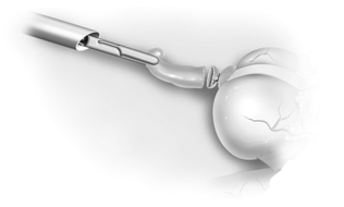
Figure 4 Pull and removal of the appendix through the left iliac fossa trocar immediately after its section, avoiding the use of an extraction bag.
Surgery was performed in 1552 patients between 2000 and 2019, 56.25% of whom were women. The mean age of the patients was 32.66years (9–93years). The patients presented with appendicitis in all stages of evolution (Table 1) from edematous, purulent, or necrotic appendicitis. n four patients, necrosis of the appendix extended to the cecum, with inflammation and perforation in two patients, requiring right colectomy, which was also completed by a totally laparoscopic route. There were no conversions to open surgery or the need for insertion of additional trocars to perform appendectomy, except in cases of conversion to right colectomy. In all cases, inspection of the entire abdominal cavity and aspiration and washing, when there were purulent collections, were possible in all quadrants through the access used. Only 84 patients underwent drainage where there was an abscess in the right iliac fossa, using Silicone Penrose drain. Antibiotic prophylaxis was used in cases of edematous appendicitis with second-generation cephalosporin. In other patients, antibiotic therapy with third-generation cephalosporin and nitroimidazole was performed. The mean length of stay was 1.74days (1–12days), with a median of 1.2 days. There were 6 rehospitalizations in the first 30days with significant abdominal pain or fever, and in 2 of these, there was a need for relaparoscopy for aspiration of purulent pelvic collection. Hyperemia and inflammation of the right iliac fossa incision developed in 74 patients (4.7%), but no case of abscess that required drainage in the incisions was noted. There was no mortality.
|
Appendicitis stages |
Number of patients |
Percentage |
|
Edematous |
821 |
52,9% |
|
Purulent |
488 |
31,4% |
|
Necrotic |
242 |
15,6% |
Table 1 Staging of appendicitis
As described by Fitz and first performed in 1886 for the treatment of acute appendicitis, appendectomy is the safest treatment for this condition at any stage of its development.8 The incisions used for laparotomic access vary widely, but the most common is the access proposed by McBurney (oblique incision in the right iliac fossa).9 The aesthetic result is quite precarious when using laparotomic incisions, whether oblique, horizontal, or vertical. Most appendectomies are performed in children and adolescents, and aesthetics is an extremely important factor in the evaluation of the appendix extraction method.10 These scars will remain for life and can change as the patient grows, often becoming extremely unsatisfactory in appearance.
The introduction of the laparoscopic route for appendectomy, described by Kurt Semm in 1982,1 brought significant aesthetic benefits since it is almost always performed with three punctures, two of which are located in different positions in the abdominal wall but may be visible when the abdomen is exposed, depending on their location. This is of particular importance when the operation is performed on female adolescents.
The access through natural holes (excluding the navel from this classification) has not had a significant evolution, and even if it can be used in the future, it will require instruments and specialized equipment, increasing the cost of the procedure, in addition to requiring highly trained staff to perform it.11 The single umbilical access technique is feasible for appendectomy and has better aesthetic appeal in relation to multiple visible incisions in the anterior abdominal wall.8,12–18
For single access, the incision needs to be larger and may become visible or may deform the patient's navel. The incision must be at least 2.5 cm long for placement of a special trocar or three conventional trocars.11,19,20 Most appendectomy indications are present in children, adolescents, and young adults. An incision of this size can determine extremely precarious aesthetic results in these cases.21,22 Descriptions of single-access case series show that an additional trocar may be required in up to 10% of cases23 or even be converted to a three-trocar technique, compromising the aesthetic aspect.15 In comparative series, it caused greater pain compared to conventional laparoscopy.24,25 The cosmetic result was considered better or comparable to the conventional method.8,12
Even if it has better aesthetic outcomes than conventional laparoscopy, the cost may be significantly higher because of the need to use special devices to introduce the instruments by single access.9,24,25 To reduce its cost, three trocars, 10 and 5mm in diameter, can be placed together through a single enlarged umbilical incision.20,26 The descriptions of this strategy led authors, in comparative series, to consider the difficult execution and aesthetic final aspect similar to laparoscopy with three portals.16,17,23 In the single-portal technique, the technical difficulty of performing the dissection and section of the appendix with conventional instruments without triangulation and in a bad position to visualize the operative field cannot be disregarded, making the procedure more risky and more difficult to apply in the appendixes that are more difficult to resect.27,28
Safety is an important aspect to consider in surgery as common as appendectomy, where even the number of procedures performed by each surgeon has implications in increasing complications, length of hospital stay, and cost of the procedure.27,29 In contrast, if performed safely and using a noninvasive method in a specialized environment, discharge rates can be achieved in less than a day in up to 90% of cases.6,30 The technique described in this study, with three portals – one umbilical and two supra-pubic – has an extremely satisfactory aesthetic result, because the scars remain hidden behind the underwear when the abdominal wall is exposed. The scar of the umbilical incision is small and may even be 5mm, not determining the deformation of the umbilical scar in any patient. The other two incisions remain hidden behind the patient’s underwear.
This technique also allows appendectomy with a privileged view of the appendix and operative instruments. It allows adequate triangulation of the instruments, resulting in safety and shorter operative time of the procedure. The use of extremely close trocars can make triangulation difficult.31 In the described technique, the trocars are located in the iliac fossa, sufficiently apart to allow adequate triangulation.
The treatment of the mesoappendix, appendix, and stump is the subject of many studies.15,32 The use of endoclips, bipolar forceps, harmonic forceps, or staplers with vascular load is described in mesoappendix release. In all or almost all these treatment methods of the mesoappendix, it remains close to the appendix, greatly increasing the volume of the operative piece and forcing its extraction in special bags, which increases the cost and time of surgery.32,33 It should be noted that the removal of the mesoappendix is merely an artifice of surgical methods, as it can remain close to the cecum without any additional risk.
Moreover, by dissecting the meso next to the appendix, the technique described here makes ligation of the appendicular artery unnecessary, avoiding the use of devices for its occlusion and reducing the risk of bleeding. We use monopolar energy at 30% of the maximum level, dissecting the mesoappendix next to the cecal appendix, where the vessels are of smaller caliber. Performed in this way, the release of the appendix does not determine the risk of significant intraoperative bleeding. There was no need for reintervention for bleeding in any of our patients. The operative specimen, being only the inflamed appendix, can be removed in almost all cases through the 10-mm trocar in the left iliac fossa, decreasing the risk of contamination of the abdominal wall during specimen removal (Figure 4).
If it is impossible for the trocar to remove the appendix, a small sterile plastic bag or an extremely low-cost piece of surgical glove is inserted through the incision of the left iliac fossa. The appendicular stump may only be sutured with 2-0 cotton thread and left exposed. Comparative studies between simple ligation and occlusion show no differences between the two methods of treatment of the appendicular stump.7 In the described technique, we performed a purse-string suture in the cecum around the appendicular stump and its invagination.
This procedure, being standardized and simplified, can be performed in any hospital environment that has a conventional laparoscopy system with the basic permanent forceps and shortens the operative time by quickly removing the piece with only one or two sutures. Because it does not use any instrument or disposable device, it has extremely low cost.34–37 It can be safely used at any age group, at any stage of appendicitis evolution, especially in morbidly obese individuals. We have systematically used this technique over the last 20years in 1552 patients who underwent surgery with low morbidity and absence of mortality. The access through three ports in all cases led to complete surgical resolution of the case, without the need for an additional trocar, attesting to its safety, efficiency, and replication. We hope with this contribution that video laparoscopic appendectomy will be more widely applied in the surgical treatment of acute appendicitis, since there is already concrete evidence about its superiority over laparotomy appendectomy, especially in more complicated cases or obese patients.
This study has several limitations. This is a retrospective study, and its results do not analyze all aspects of the patient’s evolution. It aims to demonstrate that laparoscopic appendectomy can be performed at low cost without the use of disposable material and demonstrates the safety of the application of this systemization.
The technique we have described uses three metal trocars and four permanent instruments, with a single cotton thread. The use of operating pieces, extraction bags, clips, handles, staplers or special energy, and bipolar or harmonic instruments is discontinued. Therefore, it is an extremely low-cost laparoscopic procedure. Because it allows triangulation and instrumentation in the conventional way, it is a highly safe and reproducible surgery that can be easily taught and widely used in hospitals that have conventional laparoscopic equipment. The results obtained in 1552 patients, with low morbidity and absence of mortality, attest to the safety and reproducibility of the described access.
None.
Author declares that there are no conflicts of interest.
None.

©2020 Domene, et al. This is an open access article distributed under the terms of the, which permits unrestricted use, distribution, and build upon your work non-commercially.