eISSN: 2373-6372


Letter to Editor Volume 2 Issue 3
Department of Gastroenterology, Portugal
Correspondence: Ana Filipa Ramos Ávila, Department of Gastroenterology, Hospital Divino Espírito Santo de Ponta Delgada, Avenida D Manuel I, 9500-370 Ponta Delgada, Portugal, Tel +351 296 091 000, Fax (+351) 296 203 090
Received: May 17, 2015 | Published: May 27, 2015
Citation: Ãvila F, Santos VC, Massinha P, Liberal R, Rego AC, et al. (2015) An Unusual Complication and Position 0f Pancreatic Pseudocyst:Gastric Intramural Pseudocyst.Gastroenterol Hepatol Open Access 2(3):00039. DOI: 10.15406/ghoa.2015.02.00039
Gastric intramural pseudocyst is a very rare clinical condition which may develop as a complication of chronic pancreatitis. Herein, we present the case of a 46 years-old man with a history of chronic alcoholic pancreatitis who was admitted for epigastric pain. Clinical and imaging evaluation showed a gastric intramural pseudocyst. Notably, following three months of follow-up we observed complete and spontaneous resolution of symptoms mirrored later by CT proved spontaneous drainage of the pseudocyst.Pseudocysts of the gastric wall are very rare, and the exact mechanisms leading to its formation are unknown. The drainage is indicated only to treat or prevent gastric obstruction, infection or bleeding.
Keywords: chronic pancreatitis, pancreatic pseudocyst, gastric intramural pseudocyst
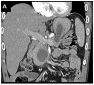
A 46-year-old male, with an alcohol-induced chronic pancreatitis, was admitted with epigastric pain. Laboratory data revealed an aspartate aminotransferase level of 98 IU/L (normal < 40), an alanine aminotransferase level of 85 U/L (normal <60) and a normal serum amylase level. Abdominal contrast enhanced computed tomography (CT) scan showed multiple pancreatic pseudocysts formations and a cystic lesion with 5,9 x 2,5 cm in diameter in the pancreatic head extending to the gastric wall which had an almost entirely intraparietal location Figure 1(A). Upper gastrointestinal endoscopy revealed bulging of the wall in the posterior portion of the gastric body, whose study by endoscopic ultrasonography confirmed the presence of a gastric intramural pseudocyst, with 55 mm in largest diameter, and with solid waste Figure 1(B). The pain resolved spontaneously, and therefore the patient was maintained on surveillance. Three months later abdominal CT showedspontaneous drainage of the intramural pseudocyst Figure 1(C).
Pancreatic pseudocysts are a common complication of both acute and chronic pancreatitis. Usually they are found within or adjacent to the pancreas, but they may also be found in other distal abdominal organs. Pseudocysts of the gastric wall are very rare, and the exact mechanisms leading to its formation are unknown.1 The possible causes of gastric intramural pseudocysts include the rupture of a pseudocyst into the gastric wall,2 the formation of a pancreatico-gastric fistula3 and the inflammation of heterotopic pancreatic tissue within the stomach.2 The drainage is indicated only to treat or prevent gastric obstruction, infection or bleeding.

Figure 1A Contrast enhanced abdominal-CT scan: multiple pancreatic pseudocyst formations and a cystic lesion with 5.9 x 2.5 cm in diameter in the pancreatic head extending to the gastric wall which had an almost entirely intraparietal location.
None.
The author declares there is no conflict of interest.

©2015 Ãvila, et al. This is an open access article distributed under the terms of the, which permits unrestricted use, distribution, and build upon your work non-commercially.