eISSN: 2373-6372


Intussusception is less common in adults than in children. Intussusception in adults is associated with an identifiable etiology in 90% of cases. Lipoma of the large intestine is rare; with a reported incidence ranging between 0.2% and 4.4%. Small Lipomas are usually asymptomatic, but giant Lipomas are often presented as abdominal pain, vomiting, diarrhea, bleeding and Intussusception. We present a case of 45-year-old man in our emergency room with intermittent abdominal cramps, rectal bleeding for eight days. Physical examination showed tenderness in the left lower abdominal quadrant. Blood found in rectal examination. Colonoscopy in two periods revealed an ulcerative mass in the proximal of descending colon. A CT scan showed a coil spring appearance highly suspicious for Intussusception of the descending-colon, a laparotomy was performed. The Intussusception was found in the descending colon, with extended left hemicolectomy, en-bloc resection was performed with end-to-end anastomosis. After surgical resection, the histo pathologic examination of the specimen showed the configuration of pedunculatedlipoma with tip ulceration, measuring 9x5x6 cm in diameter. Patient discharged on day five post-operative with good condition.
Keywords: colonic lipoma, intussusception, colonoscopy, hemicolectomy, en-bloc resection
Intussusceptions are a rare cause of bowel obstruction that accounting 1 % in adults and ileocolic valve is the most part of small bowel that involved.1 Colo colonic Intussusception accounts for 17% of confirmed intussusceptions in the literatures.2 Lymphoma and adenocarcinoma are the most leading points of Intussusception (63%) as malignant lesions.3 One of the several sub mucosal tumors is colonic Lipoma that rarely encountered in clinical practice.1 If complications of colonic lipoma such as obstruction, intussusceptions, perforation or massive hemorrhage present in patients, surgical removal of tumor is indicated.3,4 Different symptoms and presentations of colonic Lipoma in patients made it as a significant challenge in radiological confirmation and management.3–5 Here we present a 45 years old man who presented with sub-acute obstruction and rectal bleeding secondary to colo-colonic Intussusception due to a Lipoma at the splenic flexure of colon.
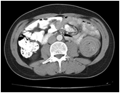
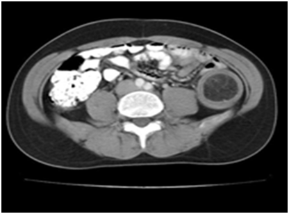
A 45 year old male patient admitted in surgical ward that complained intermittent epi gastric and left lower quadrant abdominal pain accompanied with nausea and vomiting. He has no history of medical or surgical problem. He mentioned about weight loss, moderately rectal bleeding and changing in bowel habits. In abdominal physical examination, mild tenderness over the left abdomen has been detected. Subsequent gastro endoscopy was normal. Colonoscopy was performed and find out a huge ulcerative lesion in the descending colon and pathologist reports was colitis. Computed tomography of the abdomen showed a coil spring appearance mass about 9cm in diameter in the left side of abdomen region Figures 1 & 2.

Figure 1 Computed tomography of the abdomen showed a coil spring appearance mass about 9cm in diameter in the descending colon.

Figure 2 Computed tomography of the abdomen showed a soft tissue mass (−40 till −120 Hounsfield units).
Colo-colic Intussusception was highly suspected. Second colonoscopy was done and it revealed a huge, ulcerative sub mucosal mass in the proximal portion of descending colon. Endoscopic biopsy was done and it showed a pattern of severe necrosis without evidence of malignancy. After review of above positive findings, surgical intervention was recommended. Midline laparatomy was performed; during exploration an Intussusception mass from splenic flexures to sigmoid colon was found Figure 3. An extended left hemicolectomy was done. Regional lymph node enlargement or invasion of surrounding structure was not observed. The histo pathologic examination confirmed a pedunculated Lipoma with chronic ulcer, measuring 9x5x6 cm in diameter Figure 4. After operation there, we have not any complication and patient was discharged on day five postoperative. Final pathologist report was Lipoma Figure 5.
Intussusception is a common condition in children that enlarged payer’s patches are the most cause of it but in adult, Intussusception accounts for only 5 % of all cases and 1 % of all bowel obstructions.1 The most common sites of intussusceptions are entero-entric and ileocolic.2 In adult, almost always Intussusception is secondary to pathological intra luminal lesions.2 In two thirds of adult colo-colonic Intussusception primary colonic cancers has been detected.4 Peutz-Jehger polyps, adenomas, endometriosis, previous anastomosis and lipomasare the rest cause that should concern.3 Colicky abdominal pain, rectal bleeding and bowel obstructions are the most common symptoms and palpable mass can found in 24-42% of patients.4 CT scan is the most accurate radiological diagnosis study for intussusceptions but MRI, ultrasound and barium enema have also been the other investigations.5
Colonic Lipoma can presented by abdominal pain, bleeding, constipation, perforation, bowel changes and Intussusception but in 94% of cases was asymptomatic and found incidentally during colonoscopy or surgical removed specimen.3,4,6 Diagnostic evaluations of colonic Lipoma such as barium enemas, CT scans and MRI could be complicated when Intussusception has been occurred and underlying fat necrosis can mimic malignant neoplasm’s.5 In colonoscopy visualization, characteristic mark of Lipoma is elevated normal mucosa over the tumor and fat extrusion occurs after biopsy taking.3,4,5,7
Less than 50 cases of Lipoma to us Intussusception have been reported in the literatures and just 4 cases were involved cecum-ascending colon.3 Colonic Lipoma can be treated with different options, colonoscopy and surgical procedures.8,9 Endoscopic resection of Lipoma concerned for tumors less than 2 cm, however perforation and hemorrhage can occurred during this procedure.4,8,9 Surgery has been recommended by many of the authors as the standard method of treatment for Lipoma greater than 2 cm in size.4,6,8,9 Surgical treatment includes resection, colotomy with local excision, limited colon resection, segmental resection, hemicolectomy, or subtotal colectomy; depends on the tumor size, location and presence of definitive diagnosis before operation, each kind of surgical methods that mentioned above can be used.2–4,10 In our case we do extended left hemicolectomy. The choice procedure of colonic Intussusception is resection without reduction due to the underlying malignancy as we done.10
Large colonic Lipoma with symptoms should be excised either surgically or endoscopically. Small and pedunculated Lipoma <4 cm) with normal tumor markers can undergo to endoscopic resection. Lipoma that are greater than 4 cm in size or in patients in whom malignancy cannot be ruled out should undergo segmental resection.4,9
None.
None.
The author declares that there is no conflict of interest.

© . This is an open access article distributed under the terms of the, which permits unrestricted use, distribution, and build upon your work non-commercially.