eISSN: 2473-0815


Case Report Volume 8 Issue 2
Consultant Pathologist, Maternal Perinatal National Institute, Professor, Department of Pathology, Peruvian University Cayetano Heredia, Peru
Correspondence: Jose Pereda-Garay, Consultant Pathologist, Maternal Perinatal National Institute, Professor, Department of Pathology, Peruvian University Cayetano Heredia, Peru
Received: February 06, 2020 | Published: April 15, 2020
Citation: Pereda-Garay J. Thyroid: proliferative lesions in perinatal necropsies. Endocrinol Metab Int J. 2020;8(2):48-50. DOI: 10.15406/emij.2020.08.00277
Atypical thyroid lesions have been found in a routine study of perinatal necropsies. This type of lesions means Thyroid carcinoma which is suppose result of changes in the DNA along the years. In the perinatal product, DNA changes may occur during the fecundation or abnormal DNA is transmitted by one of the parents. So families should be studied. Identification of DNA changes will prove the importance of the perinatal necropsy in the family health.
Keywords: DNA, carcinoma, necropsies, thyroid, endocrine glands, papillary carcinoma, follicular carcinoma, medullary thyroid carcinoma and anaplastic carcinoma
For long time the lack of pre-natal care and infectious diseases have been the principal cause of perinatal mortality. The solution of these problems have made evident the importance of other factors, such as geneticanomalies, in disease and mortality of all groups of age, specially perinatal cases. Malignant neoplasias are included here. In perinatal mortality the problem of neoplasias has not been studybecause cancer in the fetus and the newborn is consider rare. There are only few references in the literature of cases of this subject.1‒3 Thyroid carcinomas are an important clinical problems in adults. The objective of this paper is to illustrate different histological lesions that have been found in the thyroid during a routine survey of the archives of necropsies perinatal deaths from the National Maternal and Perinatal Institute of Lima, Peru. Some of the cases are part of multiple endocrine neoplasias, but in general even in those cases nuclear changes are more severe in thyroid that in other endocrine glands. We are presenting casesthat have been found from death because obstetric problems, so there is not a history related to the thyroid. There has not beenfounded macroscopic changes in thyroids during the dissection. They are presented as a contribution to a better understanding of the biology of this neoplasm and because their identification may be important for the family health.
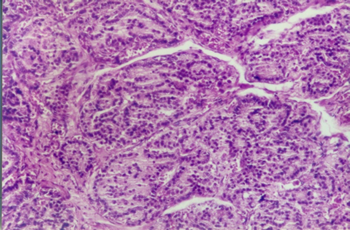
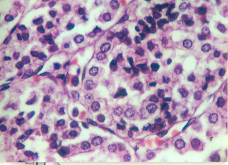
Carcinoma of the thyroid has been classified in this main histological types, papillary carcinoma, follicular carcinoma, medullary thyroid carcinoma and anaplastic carcinoma. Papillary carcinoma type. It is the more frequentlyseen in adults. We have find only one case of this type that presents acharacteristicnodule with papillary pattern (Figure 1). The aspect of nuclei of epithelial cells in no papillary areas are also atypical,looks more like those of the papillary area. Also there is a case with tall epithelial cells,that also have been related to papillary carcinoma (Figure 2).

Figure 1 Papillary nodule is seen. Appreciate that cells in no papillary areas, are different from cells of normal follicles.

Figure 2 In papillary carcinoma, a type has been describe with columnar cells.Picture showing that type of lesion.
Follicular carcinoma type.- This type was the first that call our attention because of the similarity of a section we had with the case illustrated in Ackerman´s Surgical Pathology 8th edition. Page 526published byRosai; ifwe compare the figures you can see in both of them the capsular infiltration by the tumor (Figures 3A&3B). In folliculartype,atypical nuclei with frequent mitosis are seen, and the follicles are closely together without presence of fibrovascular tissue among them (Figure 4). Hürthle cells lesion. We are not considering this lesion as a malignant tumor because we have not find the nuclear changes in this condition. But is evident that they are Hürthle cells with their typical cytoplasm filled with fine acidophilic granules due to an increased number of mitochondrias (Figure 5). We shall not comment the problem of Hürthle cell neoplasias, we mention it because in one of our cases there is an area that clearly presents glandular tissue with Hürthle cell epithelia and this fact has not been previously illustrated in the newborn.

Figure 3 This picture shows follicles with atypical epithelia and frequent mitosis. There is almost no fibro vascular stroma between the follicles.
As is stated in the beginning of this paper our intention has been to illustrate the proliferative lesions of the thyroid in perinatal cases. As it has been commented carcinogenesis is the result of multiple gene mutations along the life of the patients. In the case of the thyroid it has been describe that mutation of BRAF (proto-oncogen B-Raf) is associated to the persistence and evolution of the thyroid carcinoma more frequently papillary type. It would be interesting to find that this or other mutation are responsible for the development of the thyroid lesions we have found on the babies during the time of pregnancy. So, mutational changes have to occur very fast, more ore or less 40weeks, other possibility is that in the cases of perinatal carcinoma, the mutation is received from his parents. We have find in the literature the description of a baby with a large tumor of the neck that was an aggressive carcinoma of the thyroid resulted because a mutation at the level of exon 14 of the thyroid peroxidase of a dishormonogenetic hyperplastic goiter. The same genetic defect was found in baby´s father and her grandmother. This case shows that the mutation was in the family, and this fact is discover because a large tumor was found by clinical diagnosis. In our cases there are not macroscopic lesions; thefinding of neoplastic changes is the result of microscopic exams. The importance is that after the identification of neoplastic changes, the family can be beneficiated by the search of genetic mutation, as in the case described in the literature. If mutations are found in any of them, it will prove the importance of perinatal necropsies.We have shown the different types of proliferative lesions of the thyroid during perinatal period, in order to comment the clinical significance of the necropsies.
To Dr. Juan Rosai, who gave us permission to use a figure from his book Surgical Pathology, 8th edition. To Dr. Uriel Garcia, who read the text and made important comments that we have included.
The authors declare that there are no conflicts of interest.
None.

©2020 Pereda-Garay. This is an open access article distributed under the terms of the, which permits unrestricted use, distribution, and build upon your work non-commercially.