eISSN: 2473-0815


Case Report Volume 10 Issue 2
Ministry of Higher Education, Iraq
Correspondence: Saadi AlJadir, Ph.D., MRCP, FACE, FRCP (L), FRCP (G), Ministry of Higher Education, AlMansour, AlMutanabi, District 607, Alley 20, House 9, Baghdad, Iraq
Received: September 17, 2022 | Published: October 10, 2022
Citation: AlJadir SJS. Severe skeletal disability and abnormal biochemical tests & disease review. Endocrinol Metab Int J. 2022;10(2):47-52 DOI: 10.15406/emij.2022.10.00318
Musculoskeletal pain is the most common disorder encountered in our clinical practice that afflicts all individuals around the world and has not exempted gender, ethnicity, color, or age. The tissues which are affected are muscles, ligaments, tendons, cartilages, and bones. It can be caused by a wide range of etiologies. Chronic musculoskeletal pain causes significant morbidity and is associated with varying degrees of physical and emotional disabilities. Vitamin D deficiency has been given a major concern in the last 3 decades and has been linked with special predilection for some ethnic groups, geographical regions, high-risk groups from extreme age, social and religious customs, and most importantly sun exposure and lack of intake. We received this patient which was a young woman with good socioeconomic status and a sunny climate around the year. The patient had been referred from the Orthopedics department (late December 2013) to the Endocrine and Diabetes Clinic as having g skeletal disability and chronic pain with abnormal laboratory tests, that had eventually demonstrated secondary hyperparathyroidism with modest hypocalcemia. Clinical work-up had demonstrated hypovitaminosis D and with a musculoskeletal disorder; Osteomalacia, but the precise etiology could not be detected and some of the causes remained speculative!
Keywords: musculoskeletal disorder, osteomalacia, precise etiology, tissues
Vitamin D is a prohormone that is essential for bone and muscle health in addition to many biological functions in the human body. Some foods contain modest quantities of Vitamin (Provitamin) like dairy products, fatty fish, and mushrooms. However, the best source of Vitamin production is exposure to sunlight (ultraviolet B spectrum) for enough time during specific hours of the day and the area of skin exposed. All these factors will enable the healthy skin to synthesize the provitamins by cholesterol and ultraviolet rays. This provitamin will undergo a two-step hydroxylation in the liver (25{OH}) then the final 1-alpha hydroxylation in the kidney to produce the active vitamin (or the hormone) calcitriol. This active hormone will bind to vitamin D receptors (VDR) in many tissues and eventually exerts its biological effects. With advancing age, there will be a decline in VDR expression. Among unique biological actions is on the intestinal epithelium binding to VDRs, leading to calcium-binding protein synthesis which facilitates calcium absorption. On the other hand, the absorption of elemental oral vitamin D occurs in the duodenum and upper jejunum, therefore any pathology in this vicinity, in addition to bariatric surgery will lead to vitamin deficiency. Vitamin D production and degradation are under strict metabolic control by various feedback pathways Figure 1.
Vitamin D deficiency is a universal problem, even the sunniest regions and hot regions are not excluded Figure 2. Vitamin deficiency could be attributed to an unhealthy diet, malnutrition, malabsorption (Celiac Disease, Short Bowel), morbid obesity, and bariatric surgery. Religious restrictions obligating covering body parts, skin diseases, indoor lifestyle, special medications (for epilepsy), chronic kidney diseases, elderly and bedridden patients or living in institutions and high altitude, fear of skin cancer, extensive use of sunblock, or increased demands in pregnancy and lactation, are some of the many reasons for Vitamin D deficiency. In some countries, there is substantial awareness of this problem, therefore they have offered programmed fortification processes for many food items especially eggs and dairy products.
History
Ancient literature has told us that sun exposure was considered a remedy for physical strength and muscle building. In Greek medicine, heliotherapy was the cure for weak muscles. In addition, sun exposure was advised for better Olympic performance. Those trends were applied quite long ago before the discovery of Vitamin D. The American physician, Alfred Hess, 1922 mentioned that rachitic children with disabling muscle weakness can get tremendous improvement after direct sun exposure. Adolf Windaus was a Nobel Prize laureate in 1928 for his work about sterols that led to the discovery of the chemical structure of vitamins D2, and 7-dehydro-cholesterol. Later in the 1960s, the immediate link between muscle weakness and hypovitaminosis D was discovered Figure 3. There are enormous reports of Vitamin D deficiency from all over the regions of the world including the most developed countries like Austria, Western Europe, and the United States, besides other parts of developing countries in Asia, India, and the Middle East that have shown significant vitamin deficiency. More increasing reports and observational studies have been showing an association with many other disorders such as autoimmune diseases, T1 Diabetes Mellitus, some neoplasms, and myopathies.
The prevalence of osteomalacia among Asians from the Indian sub-continent living in the United Kingdom had been studied and many reports were available as early as the 60s, and continued later (Dunnigan et al., 1962; Ford et al., 1972a; Hodgkin et al., 1973; Polanski and Wills, 1976;). I had the privilege to work in Glasgow with one of the pioneers of those reports (MG Dunnigan) in the late 80s in Stobhill Hospital in Glasgow, Scotland where there was a big community of Pakistani and Indian individuals. The most striking findings in his study with his team had shown the best consistent variable in osteomalacia throughout the biochemical evaluations was the higher Alkaline Phosphatase among other parameters; Serum Calcium, PO4, Vitamin D 25 OH or PTH. The dressing customs of the first group are different from the Hindu women, but eventually, both groups have got significant hypovitaminosis D and Osteomalacia and interestingly documented comparable findings in their male spouses. Despite the enormous reports about the susceptibility of pregnant Asian women and their children to this disorder, the precise etiology of vitamin D deficiency in these groups remains widely open for debate.
Clinical assessment
What is the normal level of vitamin D
However, evidence indicates that optimizing parathyroid hormone level to baseline mandates Vitamin D 100 nmol/L (40 ng/ml). So, the optimal Vitamin level is more than the cut point for the deficiency.
Patient’s Lab. tests
Skeletal survey
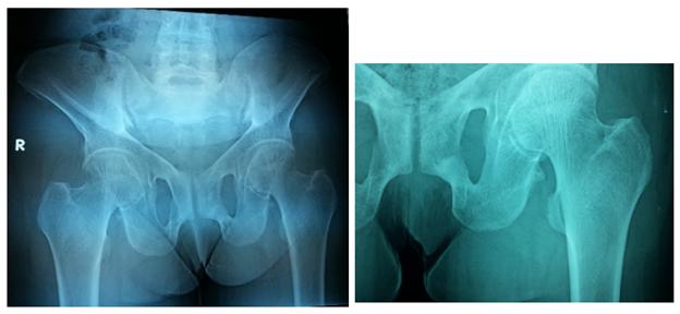
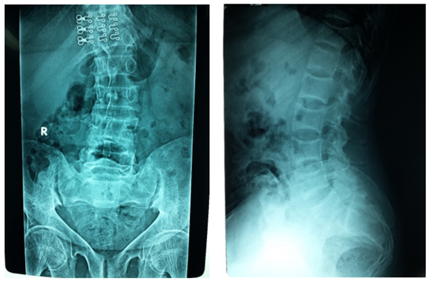
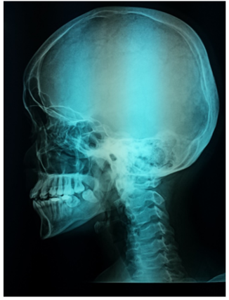
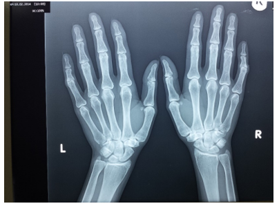
The Pelvis is markedly triradiate, with coarsening of the trabeculation of the femoral head and neck. The femoral heads have lost their normal smooth rounded outline, and are now showing relative flattening, there is a linear translucency in the sub-trochanteric region of the left femur and right inferior pubic ramus suggesting Looser’s zones Figure 4–7. The most impressive features of osteomalacia are the x-ray findings of what is considered to be diagnostic in osteomalacia. Pathologically, they are pseudo fractures except might rarely be present in Paget's disease. It appears as a thin, translucent band. Looser's zones are incomplete stress fractures that heal with callus which is poorly mineralized by calcium. They are frequently seen in the pubic bone, the necks of the femurs, and less frequently humerus and at the edge of the scapulae Figure 8.

Figure 4 Frontal X-ray of the pelvis shows Triradiate pelvis, with coarsening of the trabeculation of the femoral head and neck. The femoral heads have lost their normal smooth rounded outline and are now showing relative flattening. There is bilateral protrusion acetabula and there is a linear translucency in the subtrochanteric region of the left femur suggesting pseudo fracture or Looser’s zone.

Figure 5 Right) Frontal X-ray of LSS shows marked scoliosis and there is subchondral resorption of the right sacro-iliac joint. (Left) Lateral X-ray of LSS shows biconcave appearance of the vertebral endplates (codfish vertebrae) with marked osteopenia. There is spondylolysis of L5 on S1 and there is pathological pedicles fractures of L5. There is overall noticeable diffused osteopenia.

Figure 6 Lateral skull X-ray shows effacement (floating) of the teeth due to loss of alveolar lamina dura..

Figure 7 Frontal X-ray of both hands shows bilateral subperiosteal resorption of the radial sides of middle phalanges of middle fingers (pathognomonic of secondary hyperparathyroidism).
Vitamin D and muscle function in young population
Children with Rickets, which are characterized by chronic Vitamin D deficiency, are often manifested by substantial muscular weakness and hypotonia. On other hand, the children who harbor a rare condition called Type II Vitamin D dependent Rickets (mutation in the VDR) show muscular weakness. These observations indicate that in addition to the role of vitamin D in bone mineralization and metaphyseal closure, it also has an important role in muscular development. Older patients with osteomalacia showed proximal myopathy in both shoulder and pelvic (though most pronounced in the latter) characterized by proximal weakness, pain, unstable walking or ‘waddling gait’, and difficulty mounting stairs or rising from a sitting position or another way round.
Clinical Course of Vitamin D deficiency and Osteomalacia
Majority who have mild Vitamin D deficiencies are generally asymptomatic, or harbor nonspecific symptoms that can easily be overlooked as they might simulate many other disorders. The most pronounced symptoms of the advanced disease might be muscle pains, weakness of power, and easy fatiguability that might be relieved by rest. Osteomalacia is usually manifested by bone pains and in extreme cases easily fractured bones. The striking hallmark of the advanced stage is proximal myopathy with the pelvic girdle most severely affected. This weakness will render the patient vulnerable to falling and eventually to fracture or to immobilization. incomplete fracture or Looser’s zones are recognized by plain radiographs and might cause localized tenderness or pain on one or more bones. Other features of the disease are the gait disturbances; waddling gait, stiffness of muscles, and paresthesia (due to hypocalcemia), spinal deformities, and loss of bone density with various skeletal disabilities.
The differential diagnosis of proximal myopathy
Patient laboratory workup and effect of treatment
|
Lab Items |
Jan.14 |
April.14 |
June.14 |
|
Calcium (N 8.4-10.6 mg/dl) |
8.4 |
8.76 |
8.84 |
|
Ionized Ca (N 4.53-5.29) |
3.6 |
4.1 |
4.3 |
|
PO4 (N 2.5-4.5mg/dl ) |
2.25 |
3.33 |
3.66 |
|
Mg (N 1.41-2.29) |
- |
1.75 |
1.77 |
|
Vit.D,25(OH) |
less 7.5 |
14.2 |
21.3 |
|
AlK. Phosphatase (N 44-147 IU/L) |
209 |
172 |
164 |
|
PTH (N1.6 - 6.9 pg/ml) |
43.7 |
34.0 |
21.6 |
Patient diagnosis
Clinical features were consistent with advanced osteomalacia, due to severe Vitamin D Deficiency
Etiology could not be clearly defined.
The mode of Vitamin D administration
Vitamin D toxicity
Within the doses recommended, toxicity is rare, and cautions are only taken while treating patients with chronic granulomas (Sarcoidosis, Tuberculosis, Leprosy, Systemic fungal infection, some Lymphomas, and other rare disorders) as the inflammatory cells (Macrophages) produce 1-alpha-hydroxylase, and will produce an excess of 1,25(OH)2 D. That will lead to higher levels that might exceed 200ng/mL and serious hypercalcemia and hypercalciuria ensue.
The diagnosis
Vitamin D deficiency and Osteomalacia are usually entertained by a good history and physical examination. With appropriately selected laboratory tests and X-ray imaging of the skeleton (Skeletal survey) and it is not carried out by exclusion, as an immediate treatment even if the disease is advanced, the full recovery is usually the outcome. A point of interest in this regard is the education and awareness of vitamin D deficiency not towards the public only, but most importantly healthcare professionals whether general practitioners or specialists in regular timely methods to manage patients properly and to improve health status outcomes.
Patient’s management
Vigilance toward the etiology was kept into consideration, though we had explained to the patient that the contributing factors might be:
Recent reports have shown that Vitamin D is necessary for both bone and muscle health. The VDR is present abundantly in muscles. There is increasing evidence that Vitamin D plays an important role in counteracting muscle damage and is incorporated in regeneration. Muscle damage is characterized by a change in the architecture of muscle fibers, disruption of contractile protein, and mitochondrial dysfunction. During the insult, the VDR receptors will be enhanced in the sites of injury. As a result, this will lead to inhibition in the production of O2 radicles, which will enhance antioxidant activity and those activities that augment muscle damage which is characterized by disruption of muscle fibers, the contractile proteins, and mitochondrial dysfunction. On other hand, muscle regeneration is a very complicated process. Briefly, it involves restoration of mitochondrial function and activation of stem cells of the muscles (satellite cells). It has been shown that VDR knockdown animal models will result in decreased mitochondrial oxidative capacity and eventually energy production. It may suggest the essential role of vitamin D for mitochondrial health, and then the entire process of muscle regeneration. Therefore, it’s prudent to review the standardization of vitamin D insufficiency and the timely administration of Vitamin D analogs to maximize the role of Vitamin D. Deficiency of vitamin D results in muscle weakness and decreased speed and strength to sudden postural changes, and this will lead to falls, by 2 proposed mechanisms a) reduces inflammatory cytokines during muscle damage b) enhances myoblastic differentiation at the expense of stem cell-driven action to adipocytes Figure 9.
The author declares there is no conflict of interest.

©2022 AlJadir. This is an open access article distributed under the terms of the, which permits unrestricted use, distribution, and build upon your work non-commercially.