eISSN: 2473-0815


Opinion Volume 3 Issue 1
Primary Health Centre Belgrade, Serbia
Correspondence: Pekic Ivana, Primary Health Centre Grocka, Ljermontova 13, Belgrade, Serbia, Tel 003-816-414-310-59
Received: May 12, 2016 | Published: May 17, 2016
Citation: Ivana P. Anomalies of female sexual organs and pregnancy. Endocrinol Metab Int J. 2016;3(1):16-17. DOI: 10.15406/emij.2016.03.00037
The birth of healthy embryo is subject to many factors. It is important to detect them preconceptionally. They occur during intrauterine development and differentiation. The prevalence is 6, 7%. The can be caused by viral and parasitic diseases, the use of drugs. They are represented as the hymen without perforations, transverse buckheads and atresia vaginal, aplasia or atrezia cervical, Mayer-Rokitansky-Küster-Hauser syndrome, uterus arcuatus, uterus septus or subseptus, bicornis unicolis or bicolis, didelphus cum vagina duplex. The conse quences are amenorrea, infertility, miscarriages, premature births, low birth weight embryo. Diagnosis is made by ultrasonography, hysteroscopical, laparoscopy, MR. This individual.
We want to show the possibilities of primary health care for women for successful management of these pregnancies.
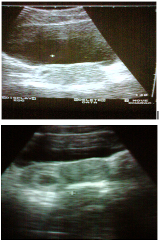
All patients were first examined in our services, when at us examinations are detected anomalities. All of them had one vagina and uterus bicornis unicolis. There were no hospitalizations. The embryos were low body weight. Conclusion/Great importance in detection of these changed has preconceptionally ultrasound examination. The course of pregnancy is caused by good connections to gynecologist at all levels of health care.1

None.
The author declares that there are no conflicts of interest.

©2016 Ivana. This is an open access article distributed under the terms of the, which permits unrestricted use, distribution, and build upon your work non-commercially.