eISSN: 2473-0815


Coexistence of acromegaly and osteosarcoma is very rare. Very few cases have been reported by the literature. Here we report the case of a 55 year-old acromegalic woman, who presented with a pathologic femoral fracture. A bone biopsy was done and an osteosarcoma was confirmed. Since the patient doesn’t have other risk factors for osteosarcoma pathology, the hypothesis is that there is a relationship between the increased rate of bone formation and the development of osteosarcoma.
Keywords: osteosarcoma, cancer, acromegaly, IGF-1, GH, insulin-dependent diabetes
IGF-1, insulin like growth factor-1; MRI, magnetic resonance imaging; GH, growth hormone
Osteosarcoma is an aggressive malignant neoplasm that affects more frequently the long bones of young people; it has a bimodal age incidence distribution, with peaks in teenagers and in adults over the age of 65.1 The peak juvenile incidence of osteosarcoma coincides with the adolescent growth spurt; the prominent factor associated is the presence of stimulated skeletal growth (insulin-like growth factor-1 (IGF-1) could promote unregulated proliferation of osteoblasts that occurs in osteosarcoma).2 As there are few reports in the literature suggesting the possible relationship between acromegaly and osteosarcoma,3 the case bellow further illustrates this coexistence. We will try through this article to discuss the possibility of causality between acromegaly and osteosarcoma.
A 55 year old woman presented a clinical feature of acromegaly for 20 years and had as antecedents: repetitive miscarriage and insulin-dependent diabetes (type 2). In 2010 the patient consulted a gynecologist for an appearance of a galactorrhoea and then she underwent a transsphenoidal surgery at Rabat’s military hospital for a sellar process approved by an MRI process. In October 2015, after a bad fall, she presented with a pain in her right hip, rebel to analgesic treatment. This pain was complicated by a pathological right femoral fracture in January 2016, which required her hospitalization in the traumatology department where she underwent a bone biopsy. The results showed that she had osteosarcoma.
Subsequently a pituitary MRI (Figure1) was done; it showed a pituitary macro-adenoma, with a superior and left lateral extension. A CT-scan exam showed multiple secondary pulmonary localizations and a laboratory evaluation confirmed the hyper-secretion of the growth hormone (GH) with a value of 27. 20ng/ml (0.01 to 3.61). The same result was found for the dosage of IGF-1 estimated to 3906.5ng/ml when female’s (41-60 yr-old) normal value ranges between 43 and 267ng/ml.
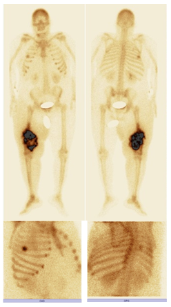
The patient was then addressed to our nuclear medicine department for a mapping of her bone lesions. Bone scintigraphy showed, in addition to the primary tumor known at the 2/3 of the right femur, a second localization to the middle arc of the 5th right rib (Figure 2).

Figure 2 99mTc-HMDP: bone scan showing, beside the primary in the 2/3 of right femur, a second localization at the middle arc of the 5th right rib.
The patient was readdressed to the endocrinology department. As a treatment, she received an injection of somatulin, and she underwent a session of Doxorubicin - Holoxan chemotherapy. Later on we were informed that she was dead at home in the end of February, after a hypoglycemic coma (reported by her daughter).
Acromegaly is caused by prolonged overproduction of GH by the pituitary gland. Secretion of GH stimulates production of IGF1 in the liver, which causes the growth of bones and other tissues in the body. Several studies suggested that patients with acromegaly have more chance to develop benign and malignant neoplasm, involving bone, brain, intestines, kidney, thyroid and especially the colon.4
Coexistence of acromegaly and osteosarcoma is an extremely rare phenomenon. There had been 5 reported patients who had these 2 diseases, the proposed hypothesis, for these cases, is that acromegaly causes the development and growth of osteosarcoma.2
It is well known that IGF-1 is a potent mitogen for a wide variety of cells, exerting its mitogenic action by increasing DNA synthesis and stimulating cell cycle progression.5 Considering the effect of GH/IGF-1 in the bone, it is established that GH is involved in the regulation of bone turnover and remodeling, probably both directly and indirectly via the production of IGF-1.6 In active acromegaly, overproduction of GH results in increased bone turnover as demonstrated by studies using biochemical markers of bone metabolism.7,8 The characteristic juvenile peak in incidence in osteosarcoma corresponds with the period of active bone development during the adolescent growth spurt. The presence of increased bone formation and resorption seen in patient with uncontrolled acromegaly could be a possible risk factor for osteosarcoma.9 The role of growth factors in osteosarcoma is supported by the case reported by Lima’s case report in 2006.2
In this case report, a 55 year old female with uncontrolled acromegaly was diagnosed with osteosarcoma in her right femur and the middle arc of the 5th right rib. Interestingly, the patient has no known risk factors for osteosarcoma which may indicate an association between this cancer and acromegaly except her high GH and IGF-1 levels that could be a risk factor for the development of her aggressive bone tumor.
Through this work, we’re describing a new association between osteosarcoma and acromegaly, which may eventually support the hypothesis that patients with uncontrolled acromegaly may be at high risk of developing osteosarcoma.
None.
The author declares there is no conflict of interest.

© . This is an open access article distributed under the terms of the, which permits unrestricted use, distribution, and build upon your work non-commercially.