eISSN: 2378-315X


Research Article Volume 2 Issue 2
1Private Practice Orthodontist, Dubai, UAE
2Dean and Professor of Orthodontics, European University College, Dubai, UAE
Correspondence: Moawia A Atia, Mamazar, Mamzar Smiles Dental and Medical Specialty Center, P.O. Box 96113, Dubai, UAE
Received: January 24, 2015 | Published: April 6, 2015
Citation: Atia MA, El-Gheriani AA, Ferguson DJ. Validity of 3 shape scanner techniques: a comparison with the actual plaster study casts. Biom Biostat Int J. 2015;2(2):64-69. DOI: 10.15406/bbij.2015.02.00026
Purpose: The study purpose was to assess the accuracy of measurements made using digital models obtained from 3Shape scanners using three different techniques:
Intraoral scanning of the patient’s mouth
Extra oral scanning of a plaster model
Model scanning with a D700 device by comparing to measurements made manually on the plaster study casts.
Methods: Measurements from the three techniques were compared to measurements made on plaster study casts.
Results: There is no significant difference in the Required Measurements as obtained from digital scanners using the three different techniques, except in the overall Bolton measurement.
Conclusion: Measurements made from digitally scanned dentitions and/or study casts compare very favourably with direct measurements from study casts. However, caution must be taken with doing the overall Bolton analysis using digitally acquired measurements.
Three-dimensional scanning of the mouth is required in a large number of procedures in dentistry such as restorative dentistry and orthodontics.1,2 The 1980s saw the introduction of the first digital intraoral scanner for dentistry by a Swiss dentist, Dr. Werner Mörmann, and an Italian electrical engineer, Marco Brandestini. Nowadays, ten intra-oral scanning devices for restorative dentistry and orthodontics have been developed, with only some being commercially available.3 Available devices include, itero (Align Technologies, San Jose, California), Lava™C.O.S (3M ESPE, Seefeld, Germany), Trios (3 shape, Copenhagen, Denmark), CEREC® AC (Sirona, Bensheim, Germany) and E4D (D4D Technologies, Richardson, Texas).4 Each of these devices has specific characteristics with the exception of the iTero and the Trios; each of the above-mentioned devices requires drying and powdering of the intraoral surfaces.5 Furthermore, individual devices are driven by various typologies of structured light sources and optical components. The CEREC® and Lava™C.O.S employ blue light-emitting diodes (LEDs) whereas laser is used as a light source in the iTero, IOS Fast Scan and E4D devices. The Trios device, which was involved in our study, works by means of confocal microscopy, with a fast scanning time; the light source provides an illumination pattern to cause a light oscillation on the object.5 For the dental practitioner, the potential benefits of using an intraoral scanner may include:
Simplification
The tasks associated with the taking of conventional impressions are no longer required. Tray selection, material mixing, cleaning and plaster pouring are all made redundant and the possibility of impression failures and model retakes is eliminated entirely.6,7
The Potential for improved accuracy
Assuming that the digital impression has been correctly obtained, material shrinkage during the curing of impression materials is removed, there can be no air bubbles, no distortion due to tray movement and no risk of there being insufficient material in the tray or inadequate adhesive.4,5
Patient comfort
The reaction from patients has been decidedly positive. The use of an intraoral scanner can be advantageous for patients with a pronounced gag reflex or with a cleft lip and palate,8 and for those who are at risk of aspiration or respiratory distress during the taking of a traditional dental impression.6,7
Efficiency and convenience
Digital workflow may improve treatment planning (with a corresponding efficiency benefit), and assist in the development of new production methods and treatment concepts.9,10 Data storage and retrieval is also facilitated,9,11 while information which is stored electronically can be shared more readily, both among professionals and also between practitioners and patients.12,13
In 2009, the accuracy of digital models produced by means of a model scanner was evaluated in a systematic review by assessing the agreement of measurements taken from digital and plaster models.14,15 It was concluded that digital models offer a high degree of validity, and measurement differences are likely to be clinically acceptable. To date only one study (which was limited in terms of its scope and the breadth of the evidential sample used), has assessed the accuracy of the various techniques for taking digital models (intraoral and extra oral).13
The aim of this study was therefore to further assess the accuracy and reliability of using digital models obtained from 3Shape scanners using three different techniques:
The validity of each of these techniques was assessed with reference to the gold standard manual measurements on plaster study casts (Figure 1).

Figure 1
The sample used in this study consisted of 40 patients seeking orthodontic treatment in the postgraduate Orthodontic Clinic at the European University College (EUC), Dubai, UAE. The study was approved by the IRB of the EUC, all subjects consented to start, in the case of minors, parents signed consent forms and minors gave their assent. Digital images for the upper and lower arches were taken with an intraoral scanning device. Duplicate impressions were then immediately taken by alginate, resulting in eighty corresponding dental stone models.
The following selection criteria were used:
Impression of the maxilla and mandible were taken using cavex colour change alginate (cavex Holland) and stainless steel impression trays (M+W-Rim lock, Germany). The impressions were disinfected and poured with elite dental stones.16 The plaster models were measured using a vernier digital calliper with an accuracy of 0.01 millimetres (mm), in a bright room and without magnification. Examiners conducted all the measurements after an initial training period. The measurements obtained (the “Required Measurements”) were as follows.17-21
All scans were recorded using a Trios-3shape intraoral scanner and a D700 extra oral scanner by the same examiner (MA) in a predetermined order.21 Intraoral Scanning with Trios started with the most distal tooth in the third quadrant continuing to the anterior teeth.22 Next, the fourth quadrant was scanned, again beginning with the most distal tooth. Scanning of the maxilla started with the most distal tooth in the second quadrant and ended at the central incisor. The first quadrant was recorded starting with the most distal tooth. When both intraoral scanning and extra oral model scanning are applied, the manufacturer’s instructions (as supplied with the scanner) were taken into account and adhered to each tooth was scanned from its buccal and lingual sides by placing the camera at an angle of forty-five degrees to the tooth axis (or as close to such angle as was possible). After scanning, the electronic files were transformed into digital models by means of Orthoanalyzer3 shape software.
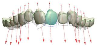
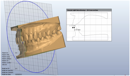
The Required Measurements were taken from the occlusal view in order to provide better visibility. For ease and accuracy of measurements, the images could be enlarged on screen and rotated as needed, using the relevant software features (Figure 2 and Figure 3). Three examiners working independently – one senior orthodontic resident as the primary examiner (MA) and two licensed orthodontists each with a minimum of three years orthodontic experience as secondary examiners – recorded the measurements of the plaster and digital models.23 Each plaster and digital model was measured twice by the primary examiner and once by each secondary examiner.

Figure 2 Measurements of mesiodistal width of upper teeth using the Orthoanalyzer 3 shape software, as shown from the facial view.

Figure 3 Selection of section plane for overbite and over jet measurements. (Left side) Study casts can be rotated which facilitates cross-sectioning at the point of maximum over jet and overbite resulting cross-sectional over jet and overbite measurements (Right side).
Statistical tests
The data was analyzed using the SPSS statistical program for Microsoft Windows XP Professional Version. The accuracy and examiner reliability of digital measurements were assessed with a paired-samplet test to compare the mean digital and manual measurements. A one way ANOVA was preformed to detect differences among the three different techniques. Pillai’s Trace, Wilks’ Lambda, Hotelling’s Trace and Roy’s Largest Root Multivariate Tests showed that our sample size is sufficient to detect any differences between the 4 groups with a 99% confidence interval (Significance level 0.01).
The Intra examiner reliability and inter examiner reproducibility was high for all measured variable (P > 0.05).24 In determining the accuracy of the measurements in three different ways in scanning it was found that all obtained measurements from the digital models were smaller than the plaster values. However, the intra oral scanning had the smallest values obtained. A one way ANOVA did not identify any statistically significant differences for all our measurements except for overall Bolton analysis (P < 0.05). The difference was between intraoral scanning models compared to plaster, digital models with D700 and extra oral scanning models (Table 1).
Mean(SD) |
Mean Difference (Std. Error) |
|||||||||
Measurement |
Plaster |
Digital (D700) |
E (Trios) |
I (Trios) |
P vs D700 |
P vs E |
P vs I |
D700 vs E |
D700 v I |
E vs I |
Overjet |
2.78(±1.4) |
2.75(±1.4) |
2.74(±1.44) |
2.68(±1.4) |
0.03(±0.32) |
0.03(±0.32) |
0.09(±0.32) |
0.00(±0.32) |
0.06(±0.32) |
0.05(±0.32) |
Overbite |
3.31(±1.5) |
3.30(±1.5) |
3.30(±1.49) |
3.18(±1.5) |
0.01(±0.34) |
0.01(±0.34) |
0.12(±0.34) |
0.00(±0.34) |
0.01(±0.34) |
0.11(±0.34) |
Max available |
73.22(±2.4) |
73.09(±2.4) |
73.09(±2.4) |
73.05(±2.4) |
0.12(±0.54) |
0.13(±0.54) |
0.17(±0.54) |
0.00(±0.54) |
0.04(±0.54) |
0.03(±0.54) |
Max required |
71.36(±4.1) |
71.24(±4.1) |
71.21(±4.1) |
71.19(±4.1) |
0.12(±0.93) |
0.14(±0.93) |
0.16(±0.93) |
0.02(±0.93) |
0.04(±0.93) |
0.02(±0.93) |
Man available |
64.09(±2.0) |
63.95(±2.2) |
63.94(±2.2) |
63.88(±2.1) |
0.14(±0.47) |
0.15(±0.47) |
0.21(±0.47) |
0.00(±0.47) |
0.07(±0.47) |
0.06(±0.47) |
Man required |
61.14(±3.4) |
61.13(±3.4) |
61.12(±3.4) |
61.09(±3.4) |
0.01(±0.77) |
0.01(±0.77) |
0.04(±0.77) |
0.00(±0.77) |
0.03(±0.77) |
0.03(±0.77) |
Max intercanine |
33.75(±2.1) |
33.75(±2.1) |
33.74(±2.1) |
33.72(±2.1) |
0.00(±0.48) |
0.00(±0.48) |
0.03(±0.48) |
0.00(±0.48) |
0.02(±0.48) |
0.02(±0.48) |
Max intermolar |
45.77(±3.0) |
45.77(±3.0) |
45.75(±3.0) |
45.93(±3.0) |
0.00(±0.67) |
0.01(±0.67) |
0.02(±0.67) |
0.01(±0.67) |
0.02(±0.67) |
0.00(±0.67) |
Man intercanine |
25.96(±2.1) |
25.95(±2.1) |
25.95(±2.1) |
25.90(±2.1) |
0.00(±0.47) |
0.01(±0.47) |
0.05(±0.47) |
0.00(±0.47) |
0.05(±0.47) |
0.04(±0.47) |
Man intermolar |
39.99(±2.8) |
39.96(±2.8) |
39.96(±2.9) |
39.63(±3.5) |
0.02(±0.68) |
0.03(±0.68) |
0.35(±0.68) |
0.00(±0.68) |
0.33(±0.68) |
0.32(±0.68) |
Ant. Bolton. |
0.76(±0.5) |
0.76(±0.5) |
0.76(±0.5) |
0.75(±0.5) |
0.00(±0.01) |
0.00(±0.01) |
0.01(±0.01) |
0.00(±0.01) |
0.00(±0.01) |
0.00(±0.01) |
Over all Bolton. |
0.92(±0.4) |
0.92(±0.4) |
0.92(±0.4) |
0.86(±0.3) |
0.00(±0.01) |
0.00(±0.01) |
0.05(±0.01)* |
0.00(±0.01)* |
0.05(±0.01)* |
0.05(±0.01)* |
Table 1 Mean, standard deviation and mean difference all measurements made on plaster and different techniques of digital models (n=40)
Confidence Intervals 95% (lower/upper) |
||||||
Measurement |
Plaster vs D700 |
Plaster vs Extra (Trios) |
Plaster vs Intra (Trios) |
D700 vs Extra (Trios) |
D700 vs Intra (Trios) |
Extra (Trios) vs Intra (Trios) |
Overjet |
-0.088/0.944 |
-0.087/0.949 |
-0.082/0.100 |
-0.908/0.919 |
-0.852/0.975 |
-0.857/0.969 |
Overbite |
-0.976/0.999 |
-0.975/0.999 |
-0.862/1.112 |
-0.986/0.988 |
-0.874/1.100 |
-0.875/1.100 |
Max available |
-1.406/1.659 |
-1.400/1.665 |
-1.362/1.703 |
-1.526/1.539 |
-1.488/1.577 |
-1.495/1.571 |
Max required |
-2.512/2.754 |
-2.485/2.781 |
-2.464/2.802 |
-2.606/2.606 |
-2.585/2.681 |
-2.612/2.654 |
Man available |
-1.187/1.476 |
-1.180/1.483 |
-1.117/1.546 |
-1.325/1.338 |
-1.262/1.402 |
-1.268/1.395 |
Man required |
-2.161/2.191 |
-2.168/2.192 |
-2.135/2.225 |
-2.179/2.181 |
-2.146/2.214 |
-2.147/2.213 |
Max intercanine |
-1.371/1.384 |
-1.368/1.387 |
-1.342/1.414 |
-1.375/1.381 |
-1.348/1.408 |
-1.351/1.405 |
Max intermolar |
-1.894/1.904 |
-1.879/1.919 |
-1.873/1.925 |
-1.884/1.913 |
-1.878/1.920 |
-1.892/1.905 |
Man intercanine |
-1.341/1.357 |
-1.335/1.362 |
-1.289/1.408 |
-1.343/1.354 |
-1.297/1.400 |
-1.163/1.392 |
Man intermolar |
-1.912/1.963 |
-1.906/1.968 |
-1.582/1.933 |
-1.932/1.943 |
-1.607/2.262 |
-1.613/2.262 |
Ant. Bolton. |
0.031/0.034 |
0.030/0.034 |
0.023/0.043 |
0.032/0.033 |
0.023/0.041 |
0.024/0.041 |
Over all Bolton. |
0.024/0.029 |
0.023/0.029 |
0.031/0.084 |
0.026/0.027 |
0.028/0.081 |
0.028/0.089 |
*Units are represented in millimeters
*P ˂ 0.05.
The third trail of our study to figure out which way of scanning to be the most accurate compare to the direct measurements on plaster models (“gold standard”). Most of the mean differences in digital models with D700 and extra oral scanning model with Trios were similar to each other. The highest mean differences were for maxillary space required and maxillary inter molar distances (means =0.02 and 0.01mm respectively, six variables have the same mean differences in plaster models and digital. Within a confidence interval of 95% we could not prove that measurements obtained from the four methods showed significant difference from each other. All of the type of scanning had errors both in positive and negative range. Only overall Bolton showed errors in the positive range for all measurements. Statistical difference was found between intraoral scanning model with trios and other various techniques and plaster models (Table 1).
In our study we relied on the research of Meredith Quimby25 to evaluate the accuracy of the three different types of scanning. Quimby used a dentoform to evaluate the accuracy of plaster models and found it useful. Plaster models were considered in our study as the gold standard to evaluate validity only.
As mentioned under the heading “Materials and Methods” above, we attempted (as far as was possible) to eliminate any factor from our study which could be responsible for measurement inconsistencies, such as any increase in time before an impression was poured in plaster, any change in the process of scanning and recording data from the patient’s mouth and the plaster model and the examiner’s lack of familiarity with the computer-based measurement of computer-based models.5
There was no significant difference in the mean measurement error obtained from all digital models for each of the three examiners. In addition, digital models showed similar measurement errors to the plaster models.26 We can therefore conclude that digital models and plaster models are alike in terms of reproducibility, similar to the inter-examiner results of Daron R Stevens,27 which showed no statistical or clinically significant difference between tooth size measurements. In contrast, other studies such as the that of Adam H. Dowling in 2013 regarding the reproducibility of digital models, demonstrated that the contact point displacement measurement data from the digital models was more reproducible than the study cast in terms of Little’s irregularity index.28
Furthermore, Anna Margreet’s29 study also concluded that digital measurements showed better reproducibility than those made on plaster models. Where there were small measurements errors in her study in terms of reproducibility, this may be explained by the possibility to zoom in on the digital images (and therefore amplify any variation) and by the lack of any physical barriers when taking the measurements.29 Measurements of space available, space required, over jet and overbite had higher mean differences when compared between plaster models and all techniques of digital scanning, whereas the measurements taken from the three different digital devices were substantially similar. This may be explained by the relative difficulty of placing the digital calliper between the teeth on the plaster models. Daron R. Stevens and Santoro27 showed a significant difference between manual and digital measurements with respect to both tooth width and overbite. The measurements from digital models were consistently smaller than the plaster model measurements.
In this study, tooth-width measurements on digital models were generally smaller than their plaster model counterparts. However, these differences were smaller than 0.4mm and could be considered as clinically insignificant. Similar results by Mathew G Wiranto4 showed that the tooth-width measurements on the digital models taken with an intraoral scanner did not differ significantly from those on the plaster models.30 In general, tooth width measurements on digital and plaster models were similar and on those teeth which showed differences, these differences were smaller than 0.2mm. The overall and anterior Bolton ratios31 differed significantly from the gold standard.
When the mean differences on the intraoral scanning and extra oral scanning models (with Trios) were compared, insignificantly small differences were found, except in the case of the overall Bolton measurement. Moreover, there were insignificantly smaller differences when the mean differences were compared between extra oral scanning models with Trios and on the digital models with the D700.Therefore, it can be concluded that the D700 scanner is the most accurate device and that intraoral scanning with the Trios is less accurate than model scanning with Trios. Differences can be exacerbated by the intraoral condition (saliva and limited spacing).
These findings have also been supported in the research of Tabea V Flügge,13 who compared inter oral scanners and model scanning to determine their precision, which was defined as the repeatability of a measured value. Intraoral scanning with iTero was found to be less precise than model scanning with iTero. It was further concluded that extra oral scanning with the D250 was more precise than extra oral scanning with the iTero. The similarity of these results with those of our study can be explained by the fact that the image acquisition technique employed by the iTero and Trios scanners does not require the application of a scanning powder.
Any differences in findings between the general bodies of research which has been undertaken in respect of digital scanning techniques may be attributable to the different technology used to acquire the relevant data. Wicher J Van der Meer5 found the Lava™ C.O.S scanner to be more accurate than the iTero and CEREC® AC scanners. He suggested that the different light sources used in these devices and their registration of the 3D images and the rest of the processing procedure may have been responsible for this difference.
Several studies have been conducted to test the accuracy and reproducibility of digital models, either by means of direct intraoral scanners or by the indirect scanning of plaster models. The question remains open as to whether the scanner impression and the bite registration has a significantly different intra arch relationship such as overbite,32 over jet and occlusion contact or if treatment plans produced with impression scanning vary substantially from treatment plans produced with plaster models.33
The findings of the study are:
The Authors thank Mr. Gafar Ouda, Mr. Peter J Norries and Dr. Laith Makki for their assistance and cooperation in this research.

©2015 Atia, et al. This is an open access article distributed under the terms of the, which permits unrestricted use, distribution, and build upon your work non-commercially.
2 7