Advances in
eISSN: 2373-6402


Research Article Volume 4 Issue 3
1School of Life Sciences, Jawaharlal Nehru University, India
2Department of Botany, University of Delhi, India
3Department of Bioscience and Biotechnology, Banasthali University, India
4Department of Plant Science, Central University of Jammu, India
5Department of Botany, University of Delhi, India
Correspondence: Neera Bhalla Sarin, School of Life Sciences, Jawaharlal Nehru University, New Delhi-110067, India, Tel +91 11 26704523, Fax +910-11-26742558
Received: January 31, 2016 | Published: August 31, 2016
Citation: Rustagi A, Shekhar S, Kumar D, et al. Genetic fidelity of in vitro cultures of an elite Indian Musa (Aa) variety Matti. Adv Plants Agric Res. 2016;4(3):292-296. DOI: 10.15406/apar.2016.04.00141
An efficient, simple, and rapid regeneration system for micro propagation of an elite Indian Banana variety Matti has been established. Shoot tip explants were regenerated on Murashige and Skoog (MS) medium supplemented with different concentrations of 6–benzylaminopurine (BAP) and Indole acetic acid (IAA). About 95% of the explants regenerated and produced on average about 28 shoots per explants, on medium supplemented with 100μM of BAP. The hardening of the in vitro grown plantlets showed almost 99% survivals upon transfer to soil in glass house conditions. Randomly amplified polymorphic DNA analysis of 10 randomly selected regenerated plants showed 100% uniformity and no detectable variation was observed.
Keywords: musa, clonal propagation, meristematic tissue, RAPD
BAP, N6–benzylaminopurine; RAPD, rapid amplification of polymorphic DNA; PCR, polymerase chain reaction; dNTPs, deoxyribonucleotide triphosphates; 2,4–D, 2,4–dicholorophenoxyacetic acid; IAA, indole acetic acid
Meristem and shoot tip cultures are the most widely used techniques for large–scale clonal propagation of banana.1–4 Regeneration via the culture of meristematic tissues located at or adjacent to the shoot apex and adventitious meristems and through the somatic embryogenic cell cultures has been used for genetic transformation of banana. In vitro propagation of Musa species has various advantages over traditional propagation such as high multiplication rate, physiological uniformity, availability of disease–free propagules, short harvest period and rapid propagation of newly selected hybrid cultivars that further help in distribution and conservation of germplasm. Apart from the influence of genotypes, shoot proliferation rate is also affected by the concentration and type of cytokinins used. Adenine–based cytokinins are used in several Musa species for in vitro propagation. N6–benzylaminopurine (BAP) is the most commonly preferred cytokinin.5,6 The others are isopentyladenine (2–ip),7 zeatin8 and kinetin.9 The concentration of cytokinin in the culture medium appears to be a crucial factor affecting multiplication. In our earlier communications, we showed 2,4–dicholorophenoxyacetic acid (2,4–D) and zeatin promotes development of embryogenic calli from scalps through somatic embryogenesis.10
In the present research work, an efficient regeneration protocol for micro propagation in an elite Indian Musa variety Matti using shoot tip explants has been developed. Rapid amplification of Polymorphic DNA (RAPD) is a polymerase chain reaction (PCR) based powerful technique to study the genetic diversity through random amplification of genomic DNA using arbitrary decamer primer. The expression of these markers is independent of tissue and environment and requires small samples of DNA for analysis. The genetic uniformity of the micropropagated plants has also been established using Random Amplified Polymorphic DNA (RAPD) markers.
Plant material
In vitro grown plantlets of banana variety Matti (AA) were obtained from National Bureau of Plant Genetic Resources, New Delhi.
Media and culture conditions
Murashige and Skoog (MS) medium (1962) was used for all in vitro culture experiments. The media used were supplemented with 3% sucrose and 20mgl–1 ascorbic acid and the pH was adjusted to 5.8 before autoclaving at 121°C for 18min. The media was gelled with 2.0gl–1 gelrite wherever required and all hormone supplementations were included before autoclaving. The cultures were maintained at 26±2°C under the light irradiance of 40µEm–2s–1 provided by cool white fluorescent lamps (Philips, India) at 16h/day photoperiod or under dark conditions wherever mentioned.
Preparation of explant
Shoot tips (about 1cm explant having the apical dome fully covered by 3–4 leaf primordia and 2–3mm of corm tissues beneath the apical dome) were harvested from in vitro grown plantlets under aseptic conditions and used as explants.
Induction of multiple shoots
The explants were cultured on MS medium supplemented with different concentrations of BAP (0, 10, 20, 40, 60, 80, 100, 120, 140μM) and with or without IAA (1μM) to induce compact meristematic clumps of multiple shoots. After two months, the multiple shoot clumps (MSC) were formed and dissected into smaller clumps with a scalpel and were sub cultured in fresh medium (having the same composition in which they were initiated) for further multiplication of shoots.
Regeneration of plantlets
The regeneration of plantlets was achieved by elongation of shoots. The multiple shoot clumps were sub cultured in MS medium supplemented with reduced concentration of BAP (10μM) for shoot elongation. In the propagation stage, shoot number (determined by counting all the shoots/explant after four months of their initiation) was recorded. Elongated shoots were detached from the base of the clump with a sterilized scalpel and sub cultured in MS medium supplemented with IAA (1 or 2μM) for rooting.
Hardening of the plantlets
Plantlets which were 6–8cm long with well established roots were transferred to plastic pots containing agro–peat (Prakruti Agrotech, Bangalore, India). The pots were covered with transparent polythene sheet with about 15holes in it for gaseous exchange and kept for 1–2months in a growth room maintained at 26°C ±2°C under 16h/day photoperiod and 70% relative humidity. The plantlets were irrigated with Hoagland solution.11 The plantlets were later transferred to earthen pots with 50% garden soil and 50% agro–peat and kept for 30days in the glass house before they were transferred to the nursery.
Isolation of DNA
Fresh leaves from the plants growing in the nursery as well as tissue culture raised plants were used for DNA extraction. The DNA was extracted using D N easy Plant Mini Kit supplied by M/s Qiagen (Germany). Quality and quantity of DNA preparations were checked by standard spectrophotometry according to Carlos et al.12
DNA amplification using Polymerase Chain Reaction (PCR)
Ten OPB (OPB 1 to OPB 10) series primers, procured from Microsynth, India (Table 1) were used in this study. Random Amplified Polymorphic DNA (RAPD) assays were carried out in 25µl reaction mixtures containing 50 mM KCl, Tris–HCl (pH 9.0 at room temperature), 1.5mM MgCl2, 0.2mM each of the deoxyribonucleotide triphosphates (dNTPs), 10pmoles primer, 0.5U Taq DNA polymerase (Bangalore Genei) and 50ng of genomic DNA. A control PCR tube containing all the chemicals, except the template DNA, was run with each primer to check for contamination. The DNA amplification was carried out in a PTC 100 thermocycler (Programmable Thermal Controller Techne Research Inc., USA), with the following conditions: 94°C (3min) initial denaturation, 44 cycles at 94°C (1min), 35°C (90sec), 72°C (90sec). The final extension cycle allowed an additional incubation for 5min at 72°C. At least two PCR amplifications were performed for each sample with RAPD primers to evaluate the reproducibility of the bands obtained. The amplified DNA fragments were separated in a 1.2% agarose gel using 1X TAE buffer according to Sambrook et al.,13 and stained with ethidium bromide. Gels were visualized and imaged using Gel Documentation System (Gel Doc Mega, Biosystematica, UK).
Data analysis
To confirm the genetic fidelity of the tissue culture grown plants, a comparison of RAPD profile from randomly selected 10 plants from over 100 tissue culture raised plants and a control plant (nursery grown plant) was carried out. Well resolved and consistently reproducible fragments ranging from 500bp to 2.5kb were scored as present or absent for RAPD profile. For detecting any genetic change, all the RAPD data was compared from the DNA samples extracted.
Primer (Operon code) |
Sequences (5´-3´) |
Total no. of bands |
Polymorphic bands |
Polymorphism (%) |
Band size (bp) |
OPB 01 |
GTTTCGCTCC |
0 |
0 |
0 |
0 |
OPB 02 |
TGATCCCTGG |
0 |
0 |
0 |
0 |
OPB 03 |
CATCCCCCTG |
0 |
0 |
0 |
0 |
OPB 04 |
GGACTGGAGT |
0 |
0 |
0 |
0 |
OPB 05 |
TGCGCCCTTC |
0 |
0 |
0 |
0 |
OPB 06 |
TGCTCTGCCC |
12 |
0 |
0 |
700 - 2400 |
OPB 07 |
GGTGACGCA |
5 |
0 |
0 |
750 - 2400 |
OPB 08 |
GTCCACACGG |
0 |
0 |
0 |
0 |
OPB 09 |
TGGGGGACTC |
4 |
0 |
0 |
1200 - 1800 |
OPB 10 |
CTGCTGGGAC |
0 |
0 |
0 |
0 |
Table 1 RAPD markers produced by 10 selected primers in micro propagated banana variety Matti
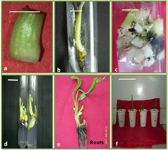
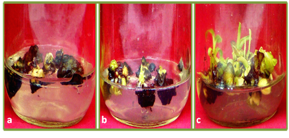
Shoot tips excised from in vitro grown plantlets (Figure 1a) cultured on MS medium supplemented with BAP at various concentrations and IAA (1 or 2 µM), showed the following developmental patterns after two months: (i) lethal blackening or lack of response (explants with pale white colour) both resulting in explant death, (ii) outgrowth of single shoots and (iii) multiplication of initial explants resulting in a cluster of small shoots (Figure 2). When shoot tips were cultured on basal MS medium, a single elongated shoot regenerated from each of the explants (Figure 1b). About 95 % of explants formed multiple shoots in MS medium supplemented with 6-BAP at concentration of 100 µM and IAA (1 µM) (Figure 1c). The average number of shoots formed per explants was 28 shoots per explant (Figure 3). Explants cultured on MS medium supplemented with 6-BAP at a concentration more than 120 µM showed blackening after one month and were discarded as they were not suitable for further propagation. Explant incubation of shoot tip showed significantly higher regeneration of shoot buds with 6-BAP (100 µM) with IAA (1µM). But the regeneration response of explants at other concentration of 6-BAP with or without IAA are significantly different from the response of 6-BAP (100 µM) with IAA (1µM). The results are similar to those of Strosse et al.14 who also reported formation of clusters of small shoots from 92% of the shoot tip explants cultured in corn shoot multiplication media (MS salts and vitamins, 30 gl-1 sucrose and 500 mgl-1 casein hydrolysate) supplemented with 100 µM 6-BAP in banana variety ‘Williams’. Transferring smaller multiple shoot clusters to lower hormonal concentration (10 µM BAP) enhanced shoot elongation. Mehta and Mohan Ram (1980) reported that higher concentration of BAP induced direct shoot regeneration while Jafari et al.15 reported that higher concentration of BAP resulted to simultaneous increment of the abnormal shoots, while as low concentration of BAP resulted into the shoot bud development from the pre-existing meristem.16,17 In contrast the present findings demonstrated that higher number of shoots (28) retrieved by 6-BAP (100 µM) with IAA (1µM) as compared to lower concentration of 6-BAP (10-80 µM) alone or in combination with IAA. The findings of present study suggested that there were no any abnormality detected at high concentration of BAP and it might be due to genotype effect. Elongated shoots derived on BAP after one month of culture (Figure 1d) were transferred to rooting medium, MS medium supplemented with IAA (1 and 2 µM) for two months. The elongated shoots have prolific rooting on the rooting medium with 3% sucrose. No difference in rooting was observed by increasing the concentration of IAA from 1 µM to 2 µM. Finally the rooted plantlets (Figure 1e) were transferred to the autoclaved agropeat for hardening (Figure 1f) and 99% of them survived the transfer to soil.
Of the 10 arbitrary RAPD primers, 3 resulted in clear and scorable amplification products. The 10 RAPD primers resulted in approximately 22 scorable band classes, ranging from 500 bp to 2500 bp in size. The number of bands for each primer varied from 4 (OPB-9) to 12 (OPB-6). No polymorphic bands were observed. Since there were no changes in the banding pattern (Figure 3-5) observed in tissue culture grown plants as compared to the nursery grown control plant, it can be concluded that the protocol for clonal propagation of banana var. Matti is successful and without any risk of genetic instability as there was no detectable variation in banana clones obtained.

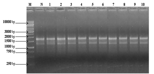
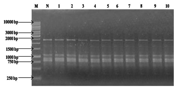
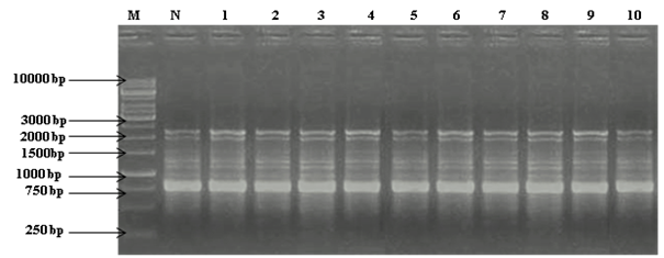
Inclusion of growth regulators in the culture medium is obligatory for inducing repeated shoot multiplication from shoot buds of banana. In this study, presence of a single cytokinin in culture medium was sufficient to induce shoot multiplication, a high rate of shoot multiplication with different combination of cytokinins has been reported,18,19,10 but there are also reports that administration of an increased concentration of growth regulators in culture medium cause variations like, suppression of shoots length leading to morphological abnormalities and genetic changes in some genotypes of banana.20,21 In the present investigation, the PCR amplification carried out by the primers selected, showed absence of any variation in the banding pattern which clearly indicates that the growth conditions and inclusion of BAP in culture medium at a concentration of 100 µM for multiple shoot formation does not induce any genetic variability in Musa variety Matti.
Absence of genetic variation using RAPD has also been reported in micropropagated shoots of Pinus thunbergii,22 chestnut rootstock hybrids ,23 Musa sp.,20,24 Almond,25 apple,26 cauliflower,27 Digitalis obscura,28 Chlorophytum arundinaceum29 and Betula pendula30 Mucuna pruriens,31 Psidium guajava.32 In all of these studies the use of RAPD proved to be useful for establishing the genetic stability. To the best of our knowledge this is the only report on the use of RAPD markers for establishing the genetic fidelity of micro propagated Indian banana variety Matti. However, soma clonal variation was also detected by RAPDs and ISSR assessment in the micro propagated banana plants, especially in Williams (AAA) Cavendish,33 Robusta (AAA) and Giant Governor (AAA).34
From this study, it can be concluded that tissue culture conditions did not induce somaclonal variation in banana variety Matti despite exposure to high level of BAP during micro propagation. Also, the protocol standardized for rapid multiplication of banana variety Matti could prove useful for commercial micro propagation due to efficient multiplication and a high degree of genetic fidelity.
In conclusion, our study describes an efficient protocol for micro propagation of an elite Indian Musa variety Matti using shoot tip explants. The high frequency shoot regeneration suggests that the standardized protocol can be employed for the germ plasm conservation. Genetic uniformity of regenerated plants suggests that tissue culture conditions did not induce variation in variety Matti despite exposure to high level of BAP during micro propagation. The efficient protocol standardized by us could greatly improve Musa germplasm handling for the purpose of clonal propagation, breeding and may also play a key role in plantain and banana improvement programme with our indigenous varieties.
Authors acknowledge the kind help of Dr. Anuradha Agrawal, National Bureau of Plant Genetic Resources, New Delhi for providing the in vitro shoot cultures of Musa variety Matti, Dr. Leela Sahijram, Indian Institute of Horticulture Research, Bangalore for providing scientific training to Dr. Anjana Rustagi. Financial assistance from UGC, CAS, UGC–RNW, DST–FIST, DST–PURSE is gratefully acknowledged. Fellowship to AR from CSIR and SS from DBT project grant are also acknowledged. Thanks to Dr. Aslam, Dr. Divya, Dr. Deepak for their valuable comments.
The author declares no conflict of interest.

©2016 Rustagi, et al. This is an open access article distributed under the terms of the, which permits unrestricted use, distribution, and build upon your work non-commercially.