MOJ
eISSN: 2374-6939


Case Report Volume 2 Issue 2
Ortho one orthopaedic specialty centre, India
Correspondence: Arun GR, Ortho one orthopaedic specialty centre, no-657, Trichy road, Singanallur, Coimbatore-641005, India, Tel 9003343337
Received: January 02, 2015 | Published: March 16, 2015
Citation: Arun GR. Concurrent shoulder dislocation with rotator cuff tear and coracoid process fracture. MOJ Orthop Rheumatol. 2015;2(2):84-86. DOI: 10.15406/mojor.2015.02.00045
Shoulder dislocation is a common consequence of athletic activity in the younger population, occurring in up to 7% of this population.1 A case concerning an adult patient with an anterior shoulder dislocation, concurrent rotator cuff tear and coracoid process fracture has not been reported in the literatura. While Bankart’s lesion with rotator cuff tear is common in older individuals, possibly due to the weakening of posterior structures in this population, young people, particularly athletes, have stronger posterior shoulder structures and are thus less prone to a rotator cuff tear when a shoulder dislocation is sustained.2 The purpose of this communication is to emphasize the need to look for rotator cuff tears and other bony injuries when evaluating shoulder dislocations in this population, and to detail the management plan.
A 55 year old gentleman came to our clinic with complains of pain and difficulty in shoulder elevation on right side. He had to suffer an episode of shoulder dislocation 2 weeks back after a road traffic accident that was reduced under anesthesia. He had significant amount of pain and was not able to elevate his right shoulder since injury.
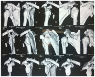
Examination revealed minimal swelling over the right shoulder. His shoulder was protected in a sling and he performed limited pendulum exercises for the first two weeks. His range of motion on the right side was significantly decreased, particularly to abduction, external rotation and internal rotation. His supraspinatus was also weak and very painful to strength testing but both anterior and posterior portions of the deltoid were intact. Neurovascular exam revealed no deficits. Preliminary x-rays showed subluxed shoulder joint. An MRI was done to evaluate the patient’s rotator cuff and glenoid labrum which showed an antero inferior labral tear, a supraspinatus, infraspinatus and subscapularis tears. A CT scan was done to check other bony pathologies. It showed a Hill sach’s lesion and coracoid process fracture which was displaced.
Examination under general anesthesia revealed shoulder instability, some crepitus with good passive range of motion. Arthroscopic evaluation showed detachment of the antero inferior labrum and full thickness supraspinatus, infraspinatus and subscapularis tear, with shallow and small Hill Sach’s lesion. No SLAP (superior labrum tear from anterior to posterior) lesion was identified. Sub acromial decompression was done. With mini open delto pectoral approach, subscapularis repair was done with a suture anchor and coracoid process fixation was done with a cortical screw. With lateral deltoid splitting approach, postero superior cuff repair was done with transosseous sutures. Shoulder joint was found to be stable after cuff repair. We did not address Bankart’s lesion because it may lead to stiffness, which is a very severe complication in elderly (Figure 1-4).

Figure 1 CT scan showing Hill Sach’s lesion, displaced coracoid process fracture and subluxed shoulder joint.

Figure 3 MRI showing signal intensity changes in supraspinatus, infraspinatus and subscapularis muscles.fatty infiltration and atrophy can be made out.
Postoperatively, the patient did well with no complications. By the time of the initial follow-up examination there had been an reduction in the level of shoulder pain. His range of motion continued to improve without apprehension but his shoulder girdle muscles did undergo noticeable atrophy. At 12 weeks, the patient progressed through physical therapy from passive to active exercises. Gradually, he achieved painless, full range of movements and a stable shoulder (Figure 5).
This is the first report of an anterior shoulder dislocation with concurrent rotator cuff tear and coracoid process fracture in the English literature to our knowledge. Coracoid process fractures constitute approximately 1% of all fractures and 2–13% of scapula fractures. Fractures are often seen on the base of the coracoid process and are generally minimally displaced and together with AC joint injuries. Indications for surgical treatment were accepted as painful nonunion, >1 cm displacement, concomitant scapula fracture on the same side and the presence of superior shoulder suspensory complex injuries.3–5
The data in elderly population is relatively different, where rotator cuff tears commonly occur in conjunction with anterior shoulder dislocation. The current literature reports incidences ranging from 35% to 86% in patients over 40 years old.2 For instance, Penvy et al.6 reported a series of 52 initial dislocations in patients older than age 40 and found that 18 (35%) had concurrent rotator cuff tears. Toolanen et al.7 reported the same incidence using ultrasound as the diagnostic test. Neviaser et al.8 used arthrograms to find an 86% incidence of rotator cuff tears in first-time anterior dislocators over the age of forty. Finally, Ribbans et al.9 found an intermediate incidence of 61% in an older population. However, Iannotti et al.10 have shown MRI to be both sensitive and specific for diagnosing labral tears, making it an attractive option when a concurrent rotator cuff tear is suspected. Further, because MRI may not differentiate between partial and full-thickness rotator cuff tears, arthroscopy is suitable for subsequent diagnostic inquiry.11 Thus, both methods were used in making this unusual diagnosis. Spormann et al.12 operated on 3 cases of isolated coracoid process fracture and obtained successful results. Again successful results were obtained from surgical treatment applied by Subramanian et al. of an isolated coracoid fracture in an unstable shoulder.13
Though conservative treatment for rotator cuff tears is often attempted first, we opted for surgery to stabilize the shoulder, which is prone for recurrent dislocation due to other associated injuries. We did a diagnostic arthroscopy first to confirm the Bankart’s lesion and massive rotator cuff tear. Later we performed a subacromial decompression and mini open deltopectoral approach to repair subscapularis tendon and internal fixation of coracoid process fracture. With mini open deltoid splitting approach, we repaired postero superior rotator cuff with transosseous sutures. The mini-open technique has become well-accepted for small and medium-sized full-thickness rotator cuff tears, particularly of the supraspinatus. It has achieved results that compare favorably to those of the traditional open procedure. For instance, Levy et al.14 reported all patients managed with mini-open repair achieved satisfactory outcomes with 80% of those being good or excellent. Further, Gumina and Postacchini have shown that surgery is superior to conservative management for dislocations and rotator cuff tears in patients over forty.
In conclusion, we present the case of a traumatic anterior shoulder dislocation with Bankart lesion, concurrent rotator cuff tear and coracoid process fracture in an adult patient. The relatively unique nature of this case derives from the patient’s age and associated injuries. Careful clinical evaluation is needed to avoid neglecting associated injuries following an episode of shoulder dislocation. Instead, the significant finding that should raise the examiner’s index of suspicion for coexistence of a rotator cuff tear or other bony injuries like coracoids process fracture is persisting severe pain after reduction of the dislocation. So, detailed clinical examination and necessary radiographic imaging can be helpful in diagnosing other associated injuries and appropriate treatment can be delivered for better functional results. While the results were excellent, no definitive treatment recommendations can be made based on this single case.

©2015 Arun. This is an open access article distributed under the terms of the, which permits unrestricted use, distribution, and build upon your work non-commercially.