Journal of
eISSN: 2379-6359


Case Report Volume 10 Issue 5
Department of Radiology, Habib Thameur Hospital, Tunisia
Correspondence: Ines Ben Hassen, Department of Radiology, Habib Thameur Hospital, Tunis, Tunisia
Received: December 12, 2018 | Published: October 1, 2018
Citation: Hassen IB, Daghfous MH. Eagle syndrome imaging: a case report. J Otolaryngol ENT Res. 2018;10(5):278?279. DOI: 10.15406/joentr.2018.10.00361
The styloid process is a conical bone formation that comes from the base of the skull to the bottom of the mastoid, the stylohyoid ligament connects the tip of the styloid process to the little horn of the hyoid bone. Eagle syndrome is a radio-clinical entity characterized by ossification of the ligament stylohyoid giving a long styloid process exceeding 30 mm in length. This syndrome is characterized by a high variability semiologic. The objective of this work is to recall the clinical presentation of Eagle syndrome in order to evoke the diagnosis and show the radiological aspect of this often misunderstood entity through a clinical case.
Eagle syndrome is a radio-clinical entity characterized by ossification of stylohyoid ligament described by Eagle in 1937.1 This entity can manifest clinical signs related to compression of neurovascular structures in the vicinity, it affects 4% of the general population of which only 4% are symptomatic. L objective of this work is to recall the clinical presentation of Eagle syndrome in order to evoke the diagnosis and show the radiological aspect of this entity.
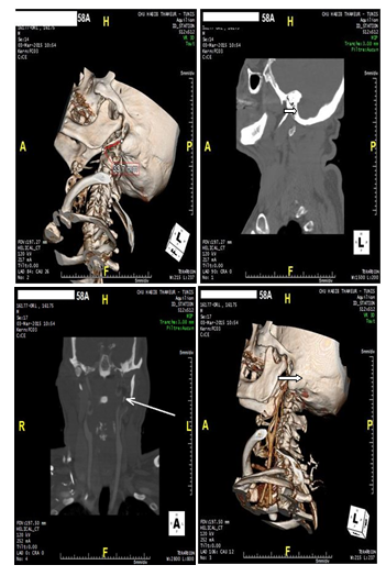
Patient 58 year old smoker, hypertensive, diabetic, dyslépidémique the history of ischemic cerebrovascular accidents for 10 years. CT angio TSA was performed as part of an exploration of atherosclerotic plaques objectified on Doppler ultrasound of TSA and accidentally shown an elongated styloid process measuring 33 last mmWG is closely related to the carotid artery external but also internal carotid artery Figure 1.

Figure 1 Reconstruction 3D styloid pro left cess; B: Sagittal; CT: elongated left styloid process
C: Coupe coronal; CT: lying left styloid process coming into contact with the carotid arteries, D: 3D reconstruction: lying left styloid process coming into contact with the carotid arteries. In réinterrogeant the patient, he says he suffered left-sided neck pain, radiating to the face. The clinical examination bilateral filling of tonsillar dimples and palpation of the styloid processes reproduced pain.
Definition
The styloid process is a conical bone formation that originates from the base of the skull to the underside of the mastoid. She heads down and inwards in the upper cervical region, the normal length varies between 20 and 25 mm in adults. The stylohyoid ligament connects the tip of the styloid process to the little horn of the hyoid bone. Eagle syndrome is a radio-clinical entity characterized by ossification stylohyoid ligament giving a long styloid process exceeding 30 mm in length.1,2
Clinical
Clinically, the Eagle syndrome is characterized by a large variability semiotic, making it impossible to identify a characteristic clinical picture Eagle distinguished three groups.
The first group is the "classic syndrome" involving a pharyngeal discomfort, neck pain, earache, foreign body sensation in the throat, dysphagia, distortion of taste and a odynophagia.
The second group is characterized by symptoms dominated by pain along the external carotid artery ride hence the name "of the external carotid syndrome."
The third group: is that of the chance discovery of ossification of stylohyoid ligament on a radiograph of the cervical spine or a panoramic radiograph in a clinically asymptomatic patient.3–6
Imaging
Standard radiology shows a continuation of the process by the styloid process of bone and a pseudo-joints appearance extending from the styloid apophysis at the small horn of hyoid (Figure 2).
Figure2.Radiopaedia elongation of the styloid process.
The dental panoramic should be interpreted with caution due to high radiographic magnification level of the patient's inclination in the radio.
CT is the reference examination before making any surgical decision.
The evaluation of the styloid process is preferably made to the scanner after contrast injection, the scanner allows for 2D reconstructions in the axis of the styloid apophysis to accurately measure its length .It also assesses the thickness of the styloid process and the relationship of the styloid apophysis with surrounding vascular structures, the tonsillar fossa and the pharynx constrictors. 3D reconstructions are most useful for evaluating the spatial relationship between the styloid process and the internal carotid artery.4–8
Treatment
The surgical treatment is based on the resection of the calcified process and the release of the neurovascular structures comprimées. Cette resection is performed either by endo-oral or externally. The operating suites are generally simple marked by the relief of symptoms. Local treatment based corticosteroid injections may be initiated in patients clinically little embarrassed or refuse the operation.10–12
Eagle syndrome is still underestimated and misunderstood by clinicians despite its frequency, palpation of tonsillar dimples and careful analysis of cervical spine radiography and panoramic radiography can evoke the diagnosis. CT is the reference examination before making any surgical decision.
None.
None.

©2018 Hassen, et al. This is an open access article distributed under the terms of the, which permits unrestricted use, distribution, and build upon your work non-commercially.