Journal of
eISSN: 2379-6359


The frontal mucocele remains a disease difficult to treat even in the era of functional endoscopic sinus surgery (FESS). Indeed, recurrence it is a typical complication of both endoscopic endonasal and transcranial approach or as a consequence of facial fracture. The modern endoscopic endonasal approach to the frontal sinus allows the marsupialization of the pathology in all those cases in which the mucocele is reached via the route of nasal drainage and is optimal when the location of the mucocele is medial. After infection or as a result of their invasion and growth, mucoceles of the frontal sinus can give rise to intracranial and orbital complications. The most versatile and used transcranial approach is considered the Sinus Fat Obliteration (SFO). We describe the case of a 47years old patient who under went FESS and OFSO for bilateral recurrence of mucocele within a mega-frontal-sinus. The lesion reached the left supra orbital region with erosion of the roof of the ipsilateral orbit. The endoscopic approach, in our experience, it is necessary in the forms of mucocele complicated by orbital medial and superior extension. Instead, in the presence of a mega-frontal-sinus, of a lateral extension of the mucocele as well and the clinical history of relapses of the disease is also indicated the transcranial surgical approach. The Sinus Fat Obliteration (SFO) minimizes the risk of complications with no need for a postoperative neurointensive unit stay compared to the intervention of cranialization. In fact the patient object of this work had a regular postoperative course and was discharged on the sixth day with excellent aesthetic results.
Keywords: frontalsinusitis, mucocele, surgery, osteotomy, fatobliteration, surgical flaps, otorhinolaryngologic surgical procedures/methods
FESS, Functional Endoscopic Sinus Surgery; SFO, Sinus Fat Obliteration

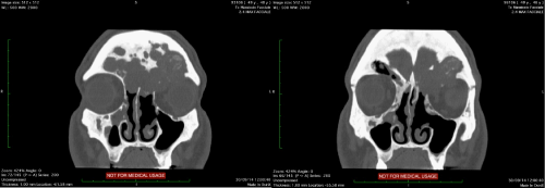
Figure 1 CT scan coronal view. The lesion reached the left supraorbital region with erosion of the roof of the ipsilateral orbit.
During surgery, it was administered to the patient perioperative broad spectrum antibiotic therapy and on awakening he did not present complications so that was conducted in the ENT Division without precautionary transfer to neurointensive unit.
During the six days of postoperative hospitalization the patient had no fever and neurological complications.
The TC postoperative control, performed after 24hours from surgery revealed no intracranial complications but the correct fat positioning in the frontal sinus.
The patient was discharged on the sixth day in excellent conditions. The subsequent endoscopic endonasal control showed no signs of recurrence.
The sinus mucocele is a disease that must be treated only by surgical therapy. The mucocele of the maxillary, ethmoid and sphenoid sinusis easily accessible by endoscopic endonasal approach. When the frontal sinus mucocele is lateral, the endonasal access is in effective to ensure the drainage function, so an extra nasal, transcranial procedure is indicated.
The fundamental objectives of SFO were the following:
CT is essential to assess the extent and localizzation of frontal mucocele , the integrity of the walls of the frontal sinus and the involvement of the other sinuses . It is the map that the surgeon uses during surgery.1
The endoscopic approach, in our experience, it is necessary in the forms of mucocele complicated by orbital medial and superior extension.
In fact, the endonasal approach allows easily reaching the muco-purulent cavity in medial and supraorbital area and getting the natural drainage through the nose. Moreover, the entire pathological mucosa present in the intranasal side can be removed and thus avoided the risk of recurrence for residues in this region.
Instead, in the presence of a mega-frontal-sinus, of a lateral extension of the mucocele as well and the clinical history of relapses of the disease is also indicated the transcranial surgical approach. It is therefore necessary an external approach to ensure the meticulous removal of the pathological mucosa. The closure of the naso-frontal duct prevents infection from nasal cavity and displacement of fat into the nose.1
So the most suitable material for the obliteration remains abdominal fat, both for its compatibility that even for the purposes of a correct radiological follow-up.
The SFO is a effective method of treatment of frontal sinus mucocele when the entire frontal sinus is not accessible by endonasal endoscopic approach.
Weber et al.,6 treated 75 primary cases, 7 revisions were necessary for a success rate of 90%.
The SFO can be considered a definitive treatment, but this procedure also has a reported long-term failure rate of up to 18%.2
Difficulties interpreting post-operative imaging can also complicate management of patients with persistent symptoms after frontal sinus obliteration. During the postoperativeperiod the MRI isindicated for closeradiological follow-up to monitor the correctpositioning of the fat oearlymucocelerelapses.6
The OFSO minimizes the risk of complications with no need for a postoperative neurointensive unit stay compared to the intervention of cranialization. In fact the patient object of this work had a regular postoperative course and was discharged on the sixth day with excellent aesthetic results.
None.
The authors declare that there are no conflicts of interest.

© . This is an open access article distributed under the terms of the, which permits unrestricted use, distribution, and build upon your work non-commercially.