Journal of
eISSN: 2373-6410


Case Report Volume 4 Issue 5
Department of Neurology, Services Institute of Medical Sciences/ Services Hospital, Pakistan
Correspondence: Sadaf Iftikhar, Department of Neurology, Services Institute of Medical Sciences/ Services Hospital, Lahore, Pakistan
Received: December 09, 2015 | Published: April 11, 2016
Citation: Iftikhar S (2016) Bilateral Thalamo-Mesencephalic Infarction: A Distinct Posterior Circulation Stroke. J Neurol Stroke 4(5): 00151. DOI: 10.15406/jnsk.2016.04.00151
Acute bilateral thalamo-mesencephalic infarction is a rare entity caused by occlusion of artery of Percheron, an uncommon anatomic variation in the brain vasculature in which a solitary arterial trunk arises from the proximal segment of either posterior cerebral artery and provides arterial supply to bilateral paramedian thalami and rostral midbrain. I herein describes the clinical presentation and neuroimaging in a patient with bilateral thalamo-mesencephalic infarction due to occlusion of the artery of Percheron.
Keywords:Artery of Percheron, Bilateral thalamo-mesencephalic infarction, Occlusion
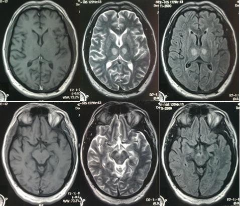
A 40-year-old non-smoker, non-alcoholic male was admitted after acute onset of stupor. On admission, his vital signs were: blood pressure 150/90mmHg, breath rate 18/min, heart rate 88/min, temperature 98.8°F. He had GCS 7/15, eyes in neutral position with pupils bilaterally equal and reactive. Oculocephalic reflex was present in all directions. He had intact bilateral corneal reflexes, symmetrical facial responses, normal gag reflex and withdrawal of all four limbs to painful stimuli. Babinski reflexes were downgoing. Vertical gaze palsy was noted with intact horizontal conjugate eye movements. All blood tests were normal including prothrombin time, activated partial thromboplastin time, activated protein C and S; thyroid profile and toxic profiles were negative. The ECG showed normal sinus rhythm and cardiac enzymes were negative. An urgent CT performed 2 hours after onset of symptoms showed no acute hemorrhage or infarction. Cerebrospinal fluid analysis was normal. MRI showed symmetrical high signal intensity on T2-weighted and FLAIR images bilaterally in the paramedian thalami and distinct V-shaped hyperintense signal in the interpeduncular fossa of midbrain (Figure 1). The posterior circulation was patent on CT angiography. Echocardiography showed no obvious valvular lesion or intra-cardiac thrombus. During hospitalization, conscious level improved markedly with fluctuant periods of somnolence while gaze palsy persisted during the follow up as well.

Figure 1 Upper: Axial T1, T2 and FLAIR MR images at the level of thalamus, showing bilateral symmetrical hyperintense signals in the paramedian nuclei.
Lower: Axial T1, T2 and FLAIR MR images at the level of midbrain, showing hyperintense signal along the pial surface of interpeduncular fossa representing the ‘V sign’.
Strokes affecting both paramedian thalami are unusual and may lead to a suspicion of an occlusion of a single arterial trunk known as the artery of Percheron.1-3 The presence of this anatomic variant must be suspected when bilateral symmetric paramedian thalamic infarcts with or without midbrain involvement, are revealed on imaging with patent basilar and posterior cerebral arteries. The clinical presentation is a triad of altered mental status, vertical gaze palsy and memory impairment.4,5 Paramedian nuclei are characterized by reciprocally activating connections with the anterior, orbitofrontal and medial prefrontal cortices through the thalamic peduncles, thus explaining the impairment in vigilance. The prominent changes in mental status may be due to the involvement of the reticular activating system as well. Presence of mydriasis and ptosis is suggestive of periaqueductal gray matter involvement. Vertical gaze palsy, as noted in our patient, is due to the involvement of the rostral interstitial nucleus of medial longitudinal fasciculus, located between the thalami and midbrain.6
These infarcts should be recognized earlier and managed accordingly and not be blamed on occlusion of multiple vascular territories or other possible etiologies such as vasculitis or infectious diseases.
None.
None.

©2016 . This is an open access article distributed under the terms of the, which permits unrestricted use, distribution, and build upon your work non-commercially.