Journal of
eISSN: 2373-633X


Research Article Volume 3 Issue 2
Department of Radiology, Pakistan Institute of Medical Sciences, Pakistan
Correspondence: Kaukab Naeem Syed, Post graduate Resident Radiology, Pakistan Institute of Medical Sciences, House no: 843, street 106 G/9-4, Islamabad, Pakistan, Tel 9.23E+11
Received: June 26, 2015 | Published: October 7, 2015
Citation: Syed KN, Zameer S, Zahoor A, et al. Diagnostic accuracy of ultrasound strain elastography for diagnosis of malignant breast lesions. J Cancer Prev Curr Res. 2015;3(2):230-233. DOI: 10.15406/jcpcr.2015.03.00072
Introduction: Current era has brought with it a dramatic climb in the incidence of malignancies especially breast carcinoma among females. It is known to be the top most diagnosed life threatening malignancy in females across the world. Such changes lead to increase need for such methods of immediate diagnosis that would lead to prompt diagnosis of the pathology, hence minimizing any delay in treatment initiation. Elastography is a technique based on the concept that hardness of the tissue increases proportionately with the pathology developing within it as compared to the normal tissue parenchyma surrounding it.
Objective: To evaluate the diagnostic accuracy of ultrasound strain elastography for determination of malignant breast lesions.
Materials and methods: Cross sectional prospective study was carried out in the Department of Radiology, Pakistan Institute of Medical Sciences Islamabad, from 1st November 2014 till 30th March 2015.Patients were selected according to predefined inclusion and exclusion criterias. Logic V5 ultrasound machine was used for evaluation of patient. Biopsy sample was then collected which was studied by consultant pathologist.
Results: Total of 43 patients was selected for the study. All were females.42 were married while 01 was unmarried. Sensitivity, specificity and diagnostic accuracy of ultrasound strain elastography was calculated to be75%, 94%, and 91% respectively.
Conclusion: Ultrasound strain elastography is an accurate, reliable and non invasive technique in the assessment of malignant breast lesions.
Keywords: breast lumps, ultrasound, strain elastography
Current era has brought with it a dramatic climb in the incidence of malignancies especially breast carcinoma among females. It is known to be the top most diagnosed life threatening malignancy in females across the world.1 There was an estimate of approximately 232,670 new cases of invasive as well as 62,570 new cases of in situ breast cancer to be diagnosed in females in the America In 2014.2 More than 1 million cases of Carcinoma breast are diagnosed each year around the world and approximately 90 thousand of these are from Pakistan.3
The above mentioned statistics for carcinoma breast are not only worrisome for the health care professionals, but also raise the need for development of such methods of immediate diagnosis that would lead to prompt diagnosis of the pathology, hence minimizing any delay in treatment initiation.
Algorithm for evaluation of palpable breast lumps is their clinical evaluation, followed by ultrasound examination and then pathological examination.4 Radiological imaging in conjunction with histopathological results play a pivotal role in early detection of clinically palpable breast lumps. In imaging, ultrasound and mammography have been relied upon significantly for the diagnosis of malignant breast lesions. However the sonographic features of benign versus malignant breast lesions cannot be accurately delineated as far as only B mode ultrasound is concerned.5,6 As Doppler has contributed to its overall sensitivity of detection of malignant breast lesions, elastography is a step ahead to add to its usefulness.
Clinically, nature of the lump is assessed by palpating its hardness. Harder the lump, greater the chances of its being malignant. Elastography is a technique based on the same concept that hardness of the tissue increases proportionately with the pathology developing within it as compared to the normal tissue parenchyma surrounding it. It is used in conjunction with the B mode ultrasound image to assess the region of interest.
The rationale of carrying out this study was to find the diagnostic accuracy of ultrasound elastography for malignant breast lesions using FNAC (Fine Needle Aspiration Cytology) and histopathology as the gold standards. Its improved sensitivity will increase the confidence level for non-invasive diagnosis of benign breast disease. It will not only lead to decline in the number of unnecessary biopsies, but will also help in preventing the avoidable delay between the definitive diagnosis of the breast lump and its treatment without waiting for the histopathological result. Histopathological examination using trucut biopsy needs on average 7-10 days for the pathologist to accurately read the slide and give out its results. On the other hand, elastography along with ultrasound is an on spot examination giving immediate conclusions. Not only is the early detection of the lump important for allying patient’s anxiety but also for its prompt treatment and to prevent its further spread.
It was prospective cross sectional study done at the department of Radiology, Pakistan Institute of Medical Sciences, Islamabad, Pakistan. It was done from 1st November 2014 till 30th March 2015. Ultrasound machine (GE Logic 5) was used for the B mode as well as ultrasound strain elastography examination of breast lumps. All female patients regardless of age were included in the study. However, the patients who were diagnosed previously or came with recurrence were excluded. Also the patients who declined trucut biopsy or who were unavailable to be followed were excluded from the study.
Informed consent was obtained from all the patients. All patients were asked about their identity and demographic profile. The data was then entered on preformed proforma.
Patients were first examined using B mode ultrasound. Suspicious lesion was then evaluated using ultrasound strain elastography by single consultant radiologist. Probe was placed centrally over the lesion. The area included was from the skin till the pectoral muscles in anteroposterior dimensions, while laterally, 5mm margins clear of the tumour were included in the field of view. Pressure was applied perpendicularly to the skin till optimal level was achieved as shown by the pressure bar in the built in software. The lesion was scored using Ueno classification as reference.7 This particular lesion was then sampled either by FNAC (fine needle aspiration cytology) or Trucut biopsy.
The cytological/histopathological results were evaluated by a single consultant pathologist who was kept blind of the results of elastographic examination.
SPSS version 17 was used to analyse data. Quantitative as well as qualitative variables were evaluated using mean, standard deviation, frequency and percentages.
A total of 43 patients were included in the study, all of whom were females. Age ranged from 16 to 45years with a mean age of 31.16+/-8.91.
Out of 43, 42 (97.6%) were married and 01(2.32%) was unmarried. Breast feeding history was positive in all married patients.12 patients (28%) had bilateral palpable breast lesions while the rest had single palpable breast lump. Axillary lymph nodes were positive in 14 patients (32.5%). Out of these, 09 (64%) were malignant looking on ultrasonographic examination with loss of fatty hilum and normal morphology. However only 06 out of these 09 patients (66.6%) had Carcinoma breast of various categorization on histopathological examination (Figure 1) (Figure 2).
Rest of 29 patients had either no lymph nodes on ultrasound examination or the fatty hilum and normal morphology was intact on B mode ultrasound.
Out of a total of 43 patients, 08 patients were labelled to be malignant while 35 patients were marked to be benign on ultrasound strain elastography. Out of these 08 malignant labelled patients, 06 were true positives when correlated with histopathological results while 02 were false positives.
Sensitivity, specificity and diagnostic accuracy of ultrasound strain elastography was calculated to be 75%, 94%, and 91% respectively.
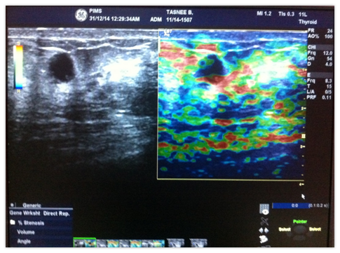
Figure 3 refers to the elastogram of a 40year old patient who was diagnosed to have score 1 lesion on strain elastography (i.e. benign) giving a BGR pattern that is typical for cystic lesions.Cyst shows layers of blue, green and red. Her histopathology report showed it to be a benign cyst.

Figure 3 40years old female; Elastography score 1; H/P Benign cyst.
BGR Pattern: It is typical for cystic lesion with layers of lue, Green and red
Figure 4 refers to the elasatogram of an 18 year old female patient whose elastography score was 2 (i.e. benign).The elastogram demonstrates a mosaic pattern of blue green and red. It turned out to be fibroadenoma on histopathological examination.

Figure 4 18years old female; Elastography score 2; H/P fibroadenoma.
The elastogram is demonstrating a mosaic of blue green and red, which represents score 2 according to Ueno classification.
Figure 5 refers to an elastogram of 42year old female patient whose elastography score was 4 .The elastogram showed the whole lesion to be of blue colour which represented its hardness. On histopathological correlation it was noticed to be invasive ductal carcinoma.
Breast cancer is one of the leading cause of female cancer deaths in the world.7 It is of utmost importance to identify the breast lump as early as possible to consider its treatment options. In the past, the breast lesions were assessed manually to look for the tissue stiffness which is related to its degree of malignant nature. Such lesions can now more reliably be evaluated by the use of ultrasound strain elastography.8 Ultrasound strain elastography is a step ahead in the diagnostic algorithm of palpable breast lesions. It not only helps in characterizing malignant breast lumps with quite an accuracy (91% diagnostic accuracy in current study), but in case of malignant lesions, the surrounding parenchyma can also be easily assessed to look for its penetration by malignant tumour growth. Hence, the treatment options for the patient can be confidently decided. Not only can it be used for breast lumps, prostate masses can also be appropriately evaluated by this recent advancement.9,10
In the current study for malignant breast lesions, it was seen that mean age of patients presenting with palpable breast lesion was 31.16+/-8.91. As compared to the literature previously published, patients in our setup presented at an earlier age group. In one study conducted by Itoh at el, mean age was calculated to be 49years while in the study carried out by Tardino et al, it came out to be 41years.11,12 According to Zhao et al, mean age was 50years in their population.13 Hence, it can be concluded that due to certain unknown circumstances, females of our population suffer from breast lumps, whether benign or malignant at much earlier age than other countries.
Diagnostic accuracy of ultrasound strain elastography in current study was calculated to be 91%. It is comparable to the studies previously conducted internationally. According to a study conducted by Hui Zhi et al.14 in japan,14 diagnostic accuracy of ultrasound strain elastography was calculated to be 88%. Also a study conducted by Liu et al.,15 accuracy of strain elastography is much higher for BIRADS 4 as well as small breast lesions.15
Combined sensitivity and accuracy of B mode ultrasound and strain elastography are even higher for detection of malignant breast lesions.15
It can hence be concluded that ultrasound strain elastography is quite accurate in identifying malignant breast lesions with a high true positive rate. It can thus reliably be used to identify malignant breast lesions. Such lesions can then be biopsied for further confirmation. However if any breast lesion turns out to be benign on ultrasound strain elastography, trucut biopsy can either be postponed or lesion can be evaluated further by using FNAC. This will not only save the patient from unnecessary financial burden of trucut biopsy but will also ally their anxiety.
None.
The authors declare there is no conflict of interests.
None.

©2015 Syed, et al. This is an open access article distributed under the terms of the, which permits unrestricted use, distribution, and build upon your work non-commercially.