Journal of
eISSN: 2373-633X


Stated purpose: Rhabdomyosarcomas are a heterogeneous group of malignant tumors representing the most common soft tissue sarcoma in lower genitourinary tract mostly occurs in infants and children in the first 2 decades of life. It is a rare and potentially deadly malignant mesenchymal neoplasia mainly originating from immature striated muscle. Rhabdomyosarcoma cells tend to appear rod-shaped, having an identifiable striated muscular differentiation with rhabdomyoblasts cells. Clinical guidelines and treatment of these tumors are very difficult because rhabdomyosarcomas behave extremely different and requires an early diagnosis and therapeutic modalities involving surgery, chemotherapy and radiation therapy.
Material & methods: In the present study, the authors report a rare case of Grade-II Penile Rhabdomyosarcoma diagnosed in a 2-year male child and have explored the histopathological literature and treatment options for this rare entity.
Conclusion: We summarized the results from the available data to the published findings which have recently reported for the proven case of grade-II rhabdomyosarcoma of the penis with treatment options for this rare entity. Considering the limited number of cases reported till now, the conclusions about treatment and prognosis are equivocal. Finally the authors concluded that penile sarcomas are rare tumors with poor prognosis when they involve deep-seated lesions.
Keywords: penile rhabdomyosarcoma, mesenchymal neoplasia, neoplasm metastasis
MRI: Magnetic Resonant Imaging; H&E: Hematoxylin & Eosin; VAC: Vincristin, Doxorubicin & Cyclophosphamide; RMS: Rhabdomyosarcoma; COG: Children’s Oncology Group; IRSG: Intergroup Rhabdomyosarcoma Study Group.
Penile sarcoma is an infrequent tumor with a very aggressive and indifferent behavior and prevalence of 0.5 to 5 cases in every 100,000 males.1 They represent only 1-2% of neoplasia in this system.2-3 Rhabdomyosarcoma is a common childhood malignancy with rare occurrence in adults and more than 50% cases are diagnosed in first 2-decades of life. These sarcomas appear to have male predominance with a ratio of 3:1.3 Normally, microscopic examination shows tumor cells arranged in sheets and lobules. Cells are pleomorphic, with round to elongated nuclei and abundant eosinophilic cytoplasm and prevalent areas of necrosis.
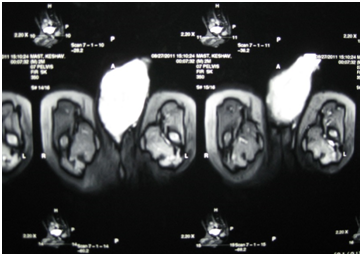
A 2-year male child presented with 2-month old history of suprapubic discomfort, dysuria and an immobile swelling over the root of the penis measuring 8.0×5.0cm in diameter. Symptoms gradually progressed and resulted in a large painless swelling over the root of the penis and enlarged bilateral testes, both measuring 4× 3.0cm in size. General physical and systemic examination was normal. The developmental milestones of the child were normal. During the local physical examination, a non-mobile hard mass was palpated which involved the root of the penis with bilateral inguinal lymphadenopathy (Figure 1). The glans penis was normal. Ultrasound of the abdomen and pelvis revealed soft tissue mass, non-homogenous in echo-pattern, 7.0×4.0×2.0 cm in size, showing internal vascularity, present within soft tissue of root of the penis inferior to pubic symphysis, inseparable from corpora cavernosa of penis. USG further revealed bilaterally enlarged testes, left and right testis measuring 4.5×3.0cm and 4.5×2.5cm in size respectively, with increased flow on color doppler and grossly altered echo-texture in bilateral scrotal sac. The right testis seems to lie at the base of the penis and compressing the corpora cavernosa. X-ray chest of the patient was normal. Magnetic resonant scan of abdomen and pelvis revealed the similar findings as on ultrasonography (Figure 2 & 3). Complete hemogram and routine blood biochemistry parameters including alpha feto protein levels of the patient were within normal limits.

Figure 1 Clinical photograph of lateral view of the giant penile growth in a 2-year male child presenting with a non-mobile hard mass measuring 8.0×5.0cm in diameter involving the root of the penis with bilateral inguinal lymphadenopathy.

Figure 2 MRI Pelvis T2W images showing bulky root of penis and a well-defined heterogenous mass lesion involving the corpracavernosa and spongiosa.

Figure 3 MRI Pelvis showing hyperintense lesion on T2W images in the root of penis suggestive of necrotic or cystic changes in it.
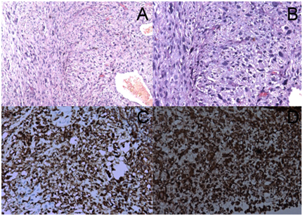
Patient underwent incisional biopsy from the penile shaft and histopathologically showed high grade soft tissue sarcoma, predominantly round cell type. The tumor was composed of pleomorphic spindle cells arranged in haphazard pattern with specifical anti-mussule antibodies (Figure 4A & 4B) and the neoplastic cells illustrated wide spread positivity for desmin and vimentin immunocytochemical stains (Figure 4C & 4D). The histopathological appearance and immunohistochemical profile of the biopsy tissue confirmed it to be a grade-II rhabdomyosarcoma of the penis.

Figure 4 Photomicrograph Hematoxylin-Eosin (H&E) stain; original magnification×100. Figure 4A & 4B: Magnification×400 and showing pleomorphic spindle cells arranged in haphazard pattern and immunohistochemical study illustrating the neoplastic cells wide spread positivity for tumor cells positive for Desmin (Figure 4C) and Vimentin (Figure 4D).
With this diagnosis, the patient underwent nine cycles of three weekly intravenous neo-adjuvant chemotherapy with VAC i.e. Inj Vincristine 7mg, Inj Doxorubicin 20 mg and Inj Cyclophosphamide 200mg. Initially patient showed good regression up to 6-cycles of VAC chemotherapy but the tumor was still unresectable and hence it was decided to give 3-more courses of VAC, however later the tumor has progressed and in view of the inoperable size of the tumor, it was decided to give second line three weekly intravenous combination chemotherapy with Inj Carboplatin 150mg and Inj Etoposide 50 mg. Despite three cycles of 2nd line chemotherapy, the disease was progressive and inoperable and the patient further developed multiple lung metastases as reported in MRI chest and abdomen in form of round lesions seen in bilateral lung peripheries. Patient later left the hospital follow-up.
Rhabdomyosarcoma is an exceedingly rare and highly malignant tumor of childhood with a typical locally invasive neoplasm that accounts for 3.8% of solid tumors in children.4-6 The diagnosis of RMS can be difficult with conventional histological techniques and extensive immunocytochemical staining is strongly recommended to establish the diagnosis. Prognostic factors include mainly lesion size (larger or smaller than 5cm), extension of invasion (superficial vs. deep-seated), complete resection of the lesion, presence or absence of metastatic disease. The described factors can help in predicting the biological behavior of sarcomas of the penis.
The main stay of treatment in pediatric patients involves multi-modal therapy that includes chemotherapy, surgery and/or radiation therapy. The best method of treatment for sarcomas is complete resection of the tumor and adjuvant chemotherapy is added after surgery in case of distant metastases.7 The main factor which is the leading reason for the poor prognosis in these patients is the fact that currently no effective response to adjuvant therapy has been obtained. Radiotherapy has been used as final control in local disease for unresectable tumors and for patients with positive margins. Diagnostic imaging plays a vital role during the close monitoring of response to therapy.2,8 Study protocols are available e.g. Children’s Oncology Group (COG), which is the successor of the Intergroup Rhabdomyosarcoma Study Group (IRSG).
Clinically, outcome for children with solid tumors have achieved significant improvement over the last three decades which may be attributed to better staging, use of risk stratification, improved local therapy and better supportive care. The Intergroup Rhabdomyosarcoma Study Group (IRSG) was formed in 1972 for methodical study of the therapy and biology of children with Rhabdomyosarcoma. During this journey span of over forty years, many chemotherapy protocols have been tried including VAC, IVA and VIE protocols (V: vincristine, A: actinomycin, E: etoposide, I: ifosfamide and C: cyclophosphamide) from IRS-I (1972–1978) to IRS-IV (1991–1997). Better after-treatment results were observed with protocol primarily consisting of VAC in terms of functional abilities and quality of life.7,8 Several studies have failed to improve outcome with the addition of chemotherapeutic agents to VAC.8 There are very few cases reported from India on Penile Rhabdomyosarcoma, the combined-modality treatment in terms of maximal patient benefit for disease control continue to be a challenge and reinforces the importance to closely monitor the systematic approach for methods of early diagnostic, disease specific targeted therapy and deep understanding of underlying biology of this disease for better outcomes in future. Finally, future directions in research may require a redefinition of risk groups to include biologic characteristics (such as PAX:FOXO1 fusion gene status) along with clinical features in risk stratification along with evaluation of response by novel imaging techniques, which may provide further data to be used to achieve this goal.8
In conclusion, we report a histopathologically proven case of grade-II rhabdomyosarcoma of the penis with treatment options for this rare entity. Considering the limited number of cases reported till now, the conclusions about treatment and prognosis are equivocal. RMS of the penis is a rare disease cured with assistance primarily by surgical intervention. The effectiveness of radiation therapy and chemotherapy is disputatious and the lack of large number of cases makes the conclusions insecure. Finally the authors concluded that penile sarcomas are rare tumors with poor prognosis when they involve deep-seated lesions. Because of the limited number of cases, currently, the best approach for treating these malignant tumors is the collaboration between the pediatric surgeon, the pathologist, the radiation and the medical oncologists in order to optimize the better treatment outcomes for the best interest of the patient.
The authors declare no conflict of interest.
None.
None.

© . This is an open access article distributed under the terms of the, which permits unrestricted use, distribution, and build upon your work non-commercially.