Advances in
eISSN: 2377-4290


Angio OCT or angiography dye less is a new method of retinal imaging based on high resolution analysis which permit to visualizate retinal and choroidal vessels. Angiography dye less we describe in this article is based on Split-spectrum amplitude-decorrelation angiography (SSADA), SSADA algorythm is based on amplitude decorrelation. We describe a new semiology of angiography dye less based on OCT and motion in retinochoroidal tissue. We present difference cases in ARMD, minimally classic choroidal new vessels, (cnv) retinal angiomatous proliferation, classic cnv, chronic cnv, and non vascular retinal epithelium detachment.
Keywords: optical coherence tomography, fluorescein angiography, angio OCT, split spectrum amplitude decorrelation, choroidal new vessel, age related macular degeneration
OCT: optical coherence tomography; FA: fluorescein angiography; SSADA: split spectrum amplitude decorrelation; cnv: choroidal new vessel; ARMD: age related macular degeneration
Optical coherence tomography (OCT) is an imaging of any tissue we can analyze with a light source,1 OCT became the gold standard for retinal disease during the last decade and take the most important place in multimodal exploration in retinal diseases. Time domain OCT (TD OCT) first, spectral domain OCT (SD OCT) and swept source OCT (SS OCT) now, are the best friends for the retinal daily practice of any ophthalmologists. OCT (SD and SS),2,3 permit an anatomic analysis of retinal tissue with now high resolution near to the μmeter, three dimensional imaging, the accuracy of these devices permit many clinical use in retinal pathology, glaucoma even in neuro ophthalmology. Angio OCT or angiography dye less is a new method of retinal imaging based on high resolution analysis which permit to visualizate retinal and choroidal vessels.
The method of angiography dye less we describe in this article is based on Split-spectrum amplitude-decorrelation angiography (SSADA). SSADA,4 based on decorrelation principles, is different than dopplers and phase approaches, it permit to analyze motions in tissue. SSADA can work with any SD and SS OCT Except on tissues motions which appears to be insignificant, the main motions are in the vessels and as a consequence, SSADA is able to perform an analysis of retinal and choroidal vascular network.
Principles of SSADA
SSADA use the natural flow as the target of the algorythm, and then, we don’t need any injection of any dye to obtain the image of retinochoroidal vascular network. The flow is detected by comparison of same acquisitions in differents times. SSADA algorythm is based on amplitude decorrelation.5 The decorrelation depends on autocorrelation (comparison of a signal with itself) and cross correlation (comparison at different time), the difference between these different signals as the variation with the time of the speckle pattern permit to detect the flow in the vessels. On the other hand than doppler techniques, based on phase analysis, SSADA aim to analyze amplitude decorrelation and is able to detect motions in axial and transverse direction, doppler OCT is only capable to measure axial flow velocity.
Clinical analysis of SSADA images
Drs Fujimoto, Lumbroso and Rispoli have made the first and very complete terminology of Angio-OCT [6], they have divided the analysis in five steps: level, reflectance, flow, morphology, texture level and flow are specifically aimed to analyze the retinochoroidal vascular network, «the level» is a basic fact for Angio OCT, this device is really capable to distinguish every level of the retinal and choroidal vasculature and to separate every stage and to review them individually, you can even scroll down the different levels to analyze the route of shunts, anastomosis, new vessels, this notion is absolutely new in retinal analysis and very informative. Angio OCT, dye less angiography, a new daily practice approach of age related macular degeneration (ARMD); «the flow» is the outright winner, the motions inside the vessels are clearly pointed out by the analyze of the vascular decorrelation signal, retinal choroidal vessels could be analyze in A 3D bulk, you can visit the network from the internal retina, to the lamina fusca over the Haller layer.
The quantification flow analysis is also a very important goal, for now it is in infancy, it is going to be a major reach in the coming year, and may be month. We can add to this really very complete classification, the notion of high resolution, the vascular images are really impressive indeed, the clearness of the picture is so good that you can examine capillaries, and the comparison with fluorescein angiography (FA) leave a gap between the two method. On the other hand Angio-OCT is not able to analyze leakage, pooling or staining like FA does, the image is static, and it is your interpretation and your enthusiasm that will be dynamic.
The process of Angio-OCT is very simple, it needs nearly the same cooperation and acquisition time than a common OCT, we can consider that this method could be extend nearly at every on Angio-OCT is motion and eye blink sensitive, but the correction of these parameters are really efficient with the news software’s, we can carry out Angio OCT in 9 cases on 10. First is a normal subject, 75 old (Figure 1), to be schematic in this presentation you can observe 4 different layers, two retinals: superficial and deep which is more medial (inner plexiform layer) and two choroidal: external retina with retinal pigment epithelium (RPE) and choriocapillaris (CC). The accuracy of the picture is real, you have a very good visualization of the peri foveolar capillaries, you discover the deep retinal vessels, densest and twisted, and then you reach the external retina avascular layer, to finish with the CC dapple-grey aspect. You can compare with OCT images to complete your diagnosis, and the level of the layer is showed on the OCT (between red and green line reported on the OCT). The first very enthusiastic Angio OCT are provided by wet complications of macular degeneration.
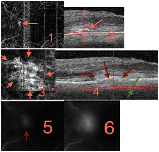
No abnormal «flow» signal is present, normal vessels are well individualized; choriocapillaris lobules are shown like clusters of half light signal. For now the best images we can obtain are 3X3 mm, possibly 6X6 mm and as a result of which the main use is centered on macula, and on ARMD In this case of subretinal new vessel which is a minimally classic choroidal new vessel7 (Figure 2) you can clearly see an abnormal flow signal pointed in red, followed by the OCT, confirmed by AF In one shot you diagnose a choroidal neovascularization and you replace AF and indocyanine green angiography (ICG). Angio OCT is also very contributive for retinal angiomatous proliferation (RAP), you can analyze your different layer and determine the course of the rap and nearly the complete morphology of the lesion. The analysis of the RAP could begin from the internal retina to the choroid In rap you observe at the retinal level, a deviation and dilation of retinal vessels in outer and medial retina (Figure 3) then you reach the anastomosis (Figure 4(1)) and the shape of the angiomatous lesion and nearly the total morphology of it (Figure 4(2,3)), the images are really better and more didactic than ICG and FA (Figure 5(1−3). The follow up of new vessels is also very interesting, with the confrontation of OCT you can easily highlight the activity of the new vessels (anastomosis, reflectance) and the regression of this activity after treatment (Figure 5−8) with sometimes very impressive images of «resting» new vessels (Figure 8,9) Angio OCT is also helpful to affirm the non vascular activity of a pigment epithelium detachment (Figure 10).

Figure 2 Minimally classic cmv: 1and 2 retinal layer, 3 et 4 CC level (green arrow) 5 FA 24s 6 FA 1’19.
Optical coherence tomography-angiography is a very efficient new method of visualization of retinochoroidal vessels; the first results in the management or ARMD are impressive and open a new area in the multimodality of the retinochoroidal investigation.
Angio OCT or angiography dye less is a new method of retinal imaging based on high resolution analysis which permit to visualizate retinal and choroidal vessels Angiography dye less we describe in this article is based on Split-spectrum amplitude-decorrelation angiography (SSADA), SSADA algorythm is based on amplitude decorrelation. We describe a new semiology of angiography dye less based on OCT and motion in retinochoroidal tissue. We present difference cases in ARMD, minimally classic choroidal new vessels, (cnv) retinal angiomatous proliferation, classic cnv, chronic cnv, and non vascular retinal epithelium detachment.
None.
Author declares that there is no conflict of interest.

© . This is an open access article distributed under the terms of the, which permits unrestricted use, distribution, and build upon your work non-commercially.