Journal of
eISSN: 2373-4426


Case Report Volume 4 Issue 2
1Departments of Paediatrics, Second University of Naples, Italy
2Paediatric Surgery Unit-Salesi Children Hospital, Italy
Correspondence: Fabiano Nino, MD, Paediatric Surgery Unit - Salesi Children Hospital, Ancona, Italy, Via Corridoni 11, 60123, Ancona, Italy, Tel +390-715-962-218, Fax -1629
Received: October 25, 2015 | Published: February 1, 2016
Citation: Papparella A, Nino F, Cobellis G (2016) Self Amputated Ovarian Cyst: An Unexpected Laparoscopic Finding. J Pediatr Neonatal Care 4(2): 00130. DOI: 10.15406/jpnc.2016.04.00130
Today laparoscopy is necessary in the diagnostic and therapeutic management of abdominal masses also in neonatal period. We report the case of a 28 days old girl admitted in our department with a prenatal diagnosis of abdominal mass. The ultrasound and CT scan revealed a 4 cm cyst in the right superior abdominal region. Instead the MRI revealed the mass localized in the right iliac region. A diagnostic laparoscopy with an open transumbilical approach was performed, that revealed an intra-abdominal 5 cm cystic formation with a thin vascular bundle. A minimal laparotomy was performed and the cyst removed. The histological examination indicated a self amputated ovarian cyst. Laparoscopy is proved to be a significant contribution in the diagnosis of abdominal masses because it allows a correct diagnosis and to plan the best management.
Keywords: laparoscopy, ovarian cyst, abdominal mass, minimal invasive surgery
Ovarian cysts are generally rare in pediatric age. They represent a heterogeneous group of conditions that range from functional (non neoplastic) ovarian cysts, to ovarian torsion, benign tumors, or even malignant and extremely aggressive neoplasm. The differential diagnosis should be made between ovarian cysts and intestinal lymphangiomas, intestinal duplication, hepatic tumors, hypersplenism, neuroblastoma ad renal masses. A precise diagnosis could be difficult for several reasons. Ovarian cysts with peduncle may be mobile generating problems in the diagnosis. Intestinal lymphangiomas and duplications show same sonographic patterns. Other abdominal masses could cause an alteration of normal anatomy through a dislocation of the surrounding organs. In these cases the common imaging techniques often cannot lead to the right diagnosis. Laparoscopy has an important role in the management of abdominal masses in infants. It guarantees a correct evaluation of anatomical relationship of the mass and its origin, and allows to plan the surgical strategy through video assisted biopsies, or the removal of the mass by laparoscopy or mini-laparotomy
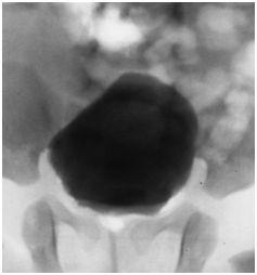
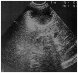
We report the case of a 28 days old girl with an antenatal diagnosis of abdominal mass. The physical examination was negative, blood sample results were within normal ranges (alpha-fetoprotein, bHCG and CA125). Urinalysis showed a small amounts of blood cells and leucocyturia, whereas the urine culture was positive for Escherichia coli (105). A micturitional cystourethrography and a complete abdominal ultrasound were performed. The micturitional cystourethrography showed a normal bladder for size and localization, with a lateral deflection of bladder imaging due to mass compression (Figure 1). The abdominal sonography showed a roundish mass with linear and exogenous margins, located in an anteromedian position compared to both the lower pole of the right kidney and the sub hepatic region. The mass was about 4x3x3 cm, and had a mixed solid -liquid echo structure (Figure 2) (Figure 3). A CT scan confirmed the presence of a 4cm cystic formation in the hepatic region but a subsequent MRI showed the mass in the right iliac fossa, and ruled out the involvement of other organs. In order to define a correct diagnosis and the subsequent surgical strategy we decided to perform a laparoscopic exploration. It was possible to identify a cystic mass in the sub-hepatic space with a diameter of 3-5cm. The mass was mobile, well delimited by the surrounding organs, with a thin long vascular pedicle (Figure 4). The exploration of the abdomen was normal and the left sided ovary and fallopian tube were normal. Although the diagnostic tests were negative, to completely rule out a neoplastic formation, the mass was totally removed through a minimal laparotomy to avoid dissemination of the fluid within the abdominal cavity in case of cyst rupture, according to the uncertain nature of che cyst. The histological diagnosis was “self amputated ovarian cyst” with necrosis and hemorrhagic fluid inside. The postoperative period was regular and the patient discharged on the second day after surgery.

Figure 1 N=57; Epidemiological distribution of the pathological fractures, traumatic fractures, and nonunion.

Figure 2 Ultrasonography: A roundish mass with linear and exogenous margins, about 4x3x3 cm with a mixed solid -liquid echo structure.
Ovarian cysts are generally rare in pediatric age. They represent a heterogeneous group of conditions that range from functional (non neoplastic) ovarian cysts, to ovarian torsion, benign tumors, or even malignant and extremely aggressive neoplasm.1,2 They could be asymptomatic or may present with symptoms referable to an acute abdomen or related to compression of adjacent organs.1 Self amputated ovarian cyst is the result of an antenatal or neonatal torsion followed by calcification and necrosis. Laparoscopy is a minimally invasive diagnostic and operative technique whereby the surgeon can treat the affected side and observe the contralateral to decide if a prophilactic pexis if it is necessary.2,3 Laparoscopy is also used in gynecology to treat the torsion of ovaries still functioning.4 In this case the diagnostic preoperative studies did not permit an adeguate diagnosis3 but laparoscopy allows a correct diagnosis, to plan the surgical intervention and to avoid a wide laparotomy. Some authors has described laparoscopic removal of ovarian cysts. But here is no general consensus because the puncture and/or rupture of the cyst could cause leakage of neoplastic cells in the abdominal cavity, even if malignancies are rare in pediatric age.5 This procedure could be carried out using lap endoscopic sack in which the cyst could be introduced to avoid chemical peritonitis in case of rupture. It was reported a non surgical treatment of self amputated ovarian cysts,6 but in our case the correct diagnosis was possible only after surgical treatment. During the last few years laparoscopy has gained consensus, becoming essential in the management of pediatric annexes pathologies. It allows an excellent visualization of all abdominal cavity and of the contralateral ovary, excellent cosmesis and a favorable outcome due to its low invasiveness.7,8 In this case laparoscopy was essential in the diagnosis and allowed the right therapeutic intervention. Its low morbidity and invasiveness proved to be a significant contribution to the treatment of this rare pathologies.
None.
The authors declare there is no conflict of interests.
None.

©2016 Papparella, et al. This is an open access article distributed under the terms of the, which permits unrestricted use, distribution, and build upon your work non-commercially.