Journal of
eISSN: 2377-4312


Mini Review Volume 12 Issue 1
Department of Veterinary Gynaecology and Obstetrics Madras Veterinary College, Tamil Nadu Veterinary and Animal Sciences University, Chennai – 600007, India
Correspondence: Akhter Rasool, Department of Veterinary Gynaecology and Obstetrics Madras Veterinary College, Tamil Nadu Veterinary and Animal Sciences University, Chennai – 600007, India
Received: August 15, 2022 | Published: January 23, 2023
Citation: Kumar RS, Rasool A, Umamageswari J, et al. Ultrasonographic evaluation of canine pyometra. J Dairy Vet Anim Res. 2023;12(1):5-6 DOI: 10.15406/jdvar.2023.12.00314
Pyometra is the most common disease found in adult intact female dogs, caused by acute or chronic suppurative bacterial infection of the uterus and is characterized by accumulation of inflammatory exudates in the uterine lumen with diverse clinic-pathological manifestation either locally or systemically. Disease is frequently noticed in adult female dog in luteal phase of estrous cycle during which progesterone level is high (progesterone sensitized uterus) and thus playing key role in pathogenesis. The preliminary diagnosis of pyometra is determined by case history, physical examination findings and laboratory test results in combination with radiography or/and ultrasonography showing a fluid-filled enlarged uterus. A late diagnosis of pyometra, when kidney failure has already occurred, may result in irreversible damage to the kidneys. Effects of sepsis and endotoxaemia can further cause multi-organ dysfunctions, but despite being a potentially life-threatening illness. In this communication, ultrasonography as an accurate procedure for the qualitative and quantitative examination and diagnosis of canine pyometra has been described.
Keywords: canine, diestrus, pyometra, ultrasound, endotoxins
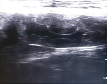
Canine pyometra is one of the most frequent reproductive organ disorders in reported in intact female dogs, particularly during the diestrus phase of estrus cycle and progesterone dominant uterus.1 Pyometra is accumulation of exudates within the uterine lumen, typically occurring during or immediately after a period of progesterone dominance. Clinical signs associated with this kind of disorder include lethargy, anorexia, polydipsia, polyuria, vomiting and unusual vaginal discharge (Kuplulu et al., 2009).2 The most accurate method of diagnosing pyometra in canines is ultrasonography.3 Both qualitative and quantitative examination is possible in diagnosing pyometra.4 In case of Pyometra, uterus will appear as distended and anechoic sacs are visualized due to pus accumulation. The advantage of ultrasonography is that it can detect the intrauterine fluid even in smaller quantity and also detect the abnormal changes in the ovaries and uterine tissue.5 Depending on the extent of uterine involvement, ultrasonographic features of pyometra may vary, the areas of uterine involvement may appear as hypoechoic or anechoic areas like if moderate involvement is there, it will appear as hypoechoic, roughly round structure ventral to ventro-lateral to the anechoic urinary bladder in transverse section. On ultrasound examination, cystic endometrial hyperplasia (CEH), which precedes pyometra, appear as small, cyst like fluid-filled regions within the endometrium (Figure 1). Similarly, transabdominal ultrasonography is effective way in diagnosing closed type of pyometra. A characteristic multiple anechoic sacculations with changes in the uterine wall thickness is visible as depicted in Figures 2–4. Therefore, ultrasound can be used as non-invasive and rapid diagnostic technique to detect the uterine pathologies like CEH and pyometra.6–10

Figure 1 Ultrasonographic view of uterus showing cystic enlargement of endometrium (CEH-Pyometra complex).

Figure 2 Ultrasonographic view of uterus showing thickened uterine wall and fluid accumulation in the lumen.
None.
None.
The authors declare no conflict of interest.

©2023 Kumar, et al. This is an open access article distributed under the terms of the, which permits unrestricted use, distribution, and build upon your work non-commercially.