Journal of
eISSN: 2377-4312


Case Report Volume 10 Issue 1
HOVET Hospital de Pequeños Animales, Práctica Privada, Madre de Dios, Perú
Correspondence: Roberto J Lope-Huaman HOVET Hospital de Pequeños Animales, Práctica Privada, Madre de Dios, Perú
Received: March 13, 2021 | Published: March 29, 2021
Citation: Lope-Huaman RJ, Huaman-Suarez RY, Curasco-Ayma A, et al. Surgical correction of a corneal-conjunctival dermoid cyst in a dog (canis lupus familiars). Clinical case. J Dairy Vet Anim Res. 2021;10(1):1-3 DOI: 10.15406/jdvar.2021.10.00303
A one and a half year old female American bully canine with the presence of chronic epiphora, with hyperemia, accompanied by bilateral uveitis, was presented for consultation. In the eye examination, a corneal choristoma was observed, from two to six o'clock (hourly nomenclature) the clinical diagnosis is compatible with a dermoid cyst with a corneo-conjunctival location in the left eye (LE). Treatment was by surgical removal of the dermoid, in surgery it was observed that the choristoma invaded the stromal layer of the cornea and encompassed the limbus and conjunctiva, with a favorable prognosis. The low frequency of appearance and location, and its surgical resolution accompanied by a prompt recovery of the cornea, makes it of interest in a clinical case, with a satisfactory result and the postoperative period passed without secondary recurrences.
Keywords: corneal dermoid, Surgical removal of the dermoid, choristoma, canine
Dermoids are choristomas, they are histologically normal tissues in an abnormal location, these being congenital malformations.1,2 Dermoid cysts are a non-neoplastic growth of skin with normal skin structure in abnormal locations. The dermoids present structural characteristics similar to the skin; They present an epidermis with variable amounts of keratin and squamous stratified cells, the dermis presents variable amounts of collagen, fibroblasts, hair follicles, sebaceous and sweat glands, it presents a hypodermis with the presence of fatty tissue.3
Dermoids occur both in man and in various species of domestic animals such as dogs, cats, parrots, cattle and horses.4 These choristomas can appear in different structures of the animal eye including that of man; such as the bulbar conjunctiva, cornea, palpebral conjunctiva, nictitating membrane.3–5 Although bilateral dermoids have been described both in man and in domestic animals, their presentation is generally unilateral. In dogs, in several studies cases of dermoids have been described mainly in the right eye.2,5,6,7
The formation of these choristomas occurs during embryonic development through defective tissue differentiation. The pathophysiology of the dermoids is still unknown. It has been described that it appears to have a genetic predisposition in some breeds of dogs, cats and cattle. Dog breeds such as the German Shepherd, Danchshunds, Golden Retrivers; in cattle, the Herefords breed seems to have a genetic predisposition.2,5,8 So far there is no classification of dermoids in dogs, but in humans there is a classification made by Ida Mann: the first type is limbic, it is the most frequent, the second type is the one that affects the cornea and can affect part of the stroma without affect Descemet's membrane and corneal endothelium, and the third type affects the entire anterior segment.6,8,9
The objective of this article was to present a clinical case of a dermoid cyst with localization lateral corneo-conjunctival between the limbus of the cornea and the sclera of the left eye (LE), that was featured in an American Bully dog and its resolution it was by surgical removal of the dermoid and its subsequent satisfactory post-surgical follow-up.
Anamnesisand physical exam
A one-and-a-half-year-old female American Bully canine patient, a one and a half year old female, presented to the HOVET Small Animal Hospital practice. For the evaluation of the presence of a type of tissue with abnormal hair in the left eye (LE), accompanied by chronic epiphora, with hyperemia, and with bilateral uveitis. On physical examination and physiological findings, respiratory rate 24 bpm, heart rate 90bpm, capillary return time 2s, pink mucosa color, SBP 120mmHg, DBP 60mmHg, and temperature 38.8˚C. They were within normal values.
Ophthalmological examination
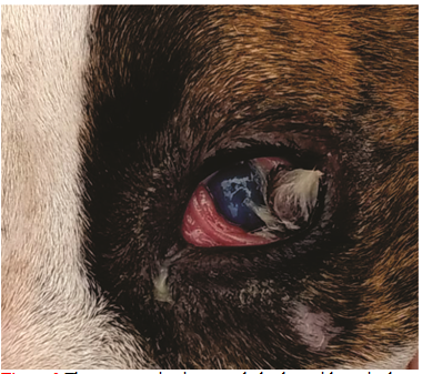
In the more detailed ocular examination, the structure of the eye was observed by magnification, it showed a mass of skin with hairs that covers 40% of the corneal surface, from two to six (hourly nomenclature) Figure 1 and Figure 2. The clinical diagnosis is compatible with a dermoid cyst with a lateral corneo-conjunctival location in the left eye (LE). It was also observed that the dermoid hairs caused irritation of the palpebral conjunctiva, these being hyperemic, also causing superficial keratitis, blepharospasm and epiphora Figure 2. To the test of test of Schirmer; They were 20 and 30mm/min in the right and left eyes, and fluorescein was negative, ruling out corneal ulceration, it was observed that there is a duct communication of the naso lacrimal duct and the consensual direct pupillary light reflections were normal in both eyes. The dog was hospitalized to undergo surgical correction of the affected structures. Previously, all the presurgical examinations such as complete blood count, kidney profile and liver profile were performed to evaluate the risk of surgery, these being in normal parameters as well as their physiological constants Table 1 and Table 2.

Figure 2 The eye examination revealed a dermoid cyst in the cornea causing superficial keratitis, blepharospasm and epiphora (LE).
|
Parameters |
Results |
Units |
Reference value |
|
Red blood cells |
5,670,000 |
Eri/uL |
5,500,000-8,200,000 |
|
Hematocrit |
48.5 |
% |
39.2-58.8 |
|
Hemoglobin |
16.16 |
g/dL |
12.7-16.3 |
|
Leukocytes |
8,500 |
Leu/uL |
6000-15000 |
|
Neutrophils |
72 |
% |
55-75 |
|
Eosinophil |
8.5 |
% |
10-Jan |
|
Lymphocytes |
eleven |
% |
1230 |
|
Platelets |
170,000 |
Plt/uL |
160000-461000 |
Table 1 Hematological parameter
|
Parameters |
Results |
Units |
Reference value |
|
Albumin |
3.2 |
mg/dL |
2.9-4.2 |
|
Bilirubin |
0.9 |
mg/dL |
0.1-0-61 |
|
Total globulin |
70 |
mg/dL |
69-120 |
|
Creatinine |
1.5 |
mg/dL |
0.5-1.6 |
|
Urea |
39 |
mg/dL |
20-40 |
|
Glucose |
89 |
Mg/dL |
69-120 |
Table 2 Blood chemistry
Surgical treatment
The anesthetic plan used consisted of the use of midazolam (benzodiazepine) (0.1mg/kgiv) as a tranquilizer and muscle relaxant, Fentanyl (opioid) (0.04mg/kgiv) was used as an analgesic and Ketamine (9mg/kg) was used as dissociative induction. kg iv) and propofol maintenance (0.51mg/kg/miniv). Infusion continues diluted in 5% glucose. At the ophthalmic level, local anesthesia drops Proparacaine hydrochloride 0.5% (Instill 2 drops) were used in the eye to obtain topical anesthesia and keep the eye open to facilitate surgery.10,11,12
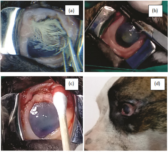
Removal of the dermoid was by surgical removal. The incision was made around the periphery of the dermoid, starting at the limit of the lateral palpebral conjunctiva, and towards the bulbar conjunctiva near the corneascleral limbus.2 The excision was continued bordering the dermoid from lateral to medial with a linear incision with the help of a curved mosquito hemostat, to achieve complete excision of the dermoid. On the other hand, it was observed that it invaded the stromal layer of the cornea and encompassed the limbus of the cornea and the sclera. All dermoid tissue was removed. LaterThis was followed by closure with a discontinuous 4-point suture. With absorbable 6-0 polyglycolic acid thread. For good post-operative management, an Elizabethan collar was put on to prevent the dog from contacting the eye (Figure 3).

Figure 3 Photograph of the same dog. (a) Dermoid cyst with lateral corneo-conjunctival location in the left eye (LE). (b) Dermoid extraction was the surgical removal. (c) Corneal surface after dermoid extraction. (d) Post-surgical ophthalmic evaluation 4 days after the intervention.
Post-operative drug treatment consisted of Cephalexin 125mg/12h. Carprofen 20mg/12h enteral route for 3 days.10 Locally, sterilized ophthalmic ointment (Healing) based on Neomycin sulfate 0.5g was applied; Gentamicin sulfate 0.3g, lidocaine hydrochloride 0.005g, L cystine 0.6g, L-Asparagine 0.22g, L-glutamine 0.4g, vitamin D2 (ergocalciferol 400000IU/g) 0.082g at a dose of 0.5cm of ophthalmic ointment in the conjunctival cul-de-sac of the affected eye every 6 hours. The duration of the treatment was for 10 days. This combination of antibiotic action and the epithelializing and tissue regeneration action helped the evolution to be satisfactory. In their weekly check-ups, the integrity of the cornea was evaluated using the Schirmer test and fluorescein, resulting negative, showing a satisfactory post-surgical recovery (Figure 4).13
According to the Ida Mann classification, the dermoid in this clinical case is of the limbal or epidular dermoid type, being the most common type of dermoid, it does not incorporate the entire cornea.11 A previous study showed that 77% of ocular dermoid cases occurred in female dogs, this coincides with our clinical case, since the dermoid was extracted from an American Bully bitch.7,8
Surgical excision (surgical excision) for resolution of ocular dermoids is consideredthe most widely used resolutive treatment of choice, with fewer post-operative complications. This is consistent with our clinical case, where we had no post-operative problems and the resolution was satisfactory.2,5,8,13
Although its presentation is generally unilateral, in dogs in several works cases of dermoids have been described mainly in the right eye. In our clinical case it was in the left eyewhich agrees with the case of a labrador.3,4
The ocular dermoid could have a hereditary pattern. Recent reports show that this may be possible. It was reported that in one litter four puppies had ocular dermoids including the father, in Herefords cattle, autosomal recessive and polygenic inheritance was observed, this was observed in a group of animals that had bilateral dermoids.3,8,13–15
The clinical case: It is of interest because its clinical presentation of the dermoid is in the left eye of an American bully dog, beingthe first report in Puerto Maldonado, on the other hand hisclinical diagnosis was evidenced by the presence of the dermoid cystof corneo-conjunctival location, In surgery, it was observed that the dermoid cyst invaded the stromal layer of the cornea and covered the limbus and conjunctiva. The reviewed literature indicates that in recent years they suggest an inherited predisposition to dermoid development and are rare in dogs. On the other hand, it is urged to continue showing in reports more similar cases at the local and national level.
None.
No potential conflict of interest was reported by the authors.
None.

©2021 Lope-Huaman, et al. This is an open access article distributed under the terms of the, which permits unrestricted use, distribution, and build upon your work non-commercially.