eISSN: 2471-0016


Case Report Volume 2 Issue 6
1Faculty of Health Sciences, University ICESI, Colombia
2School of medicine, Universidad del Valle, Colombia.
Correspondence: Liliana Fernandez T, Fundacion Valle del Lili, Avenue Simon Bolivar Av. 98 # 18-49 Tower 2 Floor 4 Consulting room 446, Cali, Colombia
Received: January 01, 1971 | Published: September 6, 2016
Citation: Fernandez TL, Sua LF, Munoz LS, et al. A rare cause of massive hemoptysis: bilateral endobronchial lesions secondary to metastatic gestational choriocarcinoma in a post-menopausal woman. Int Clin Pathol J. 2016;2(6):131–133. DOI: 10.15406/icpjl.2016.02.00058
Massive hemoptysis is a challenge in the patient with neoplastic lung disease due to its high mortality and the demand of a multidisciplinary approach. Its management entails intensive care, interventional pulmonology, hemodynamics, radiology and surgery. We present the case of a patient with obstructive endobronchial lesions and severe hemoptysis diagnosed as metastatic gestational choriocarcinoma, who was successfully treated with endoscopic intervention and chemotherapy.
Keywords: hemoptysis, choriocarcinoma, SRY, gene amplification
Massive hemoptysis is a fearsome complication of lung cancer which is found in 7-10% of the patients. It is more frequent in the cases of central lesions in the airway and can lead the patient to severe compromise of the general status, respiratory failure and death, with a mortality rate estimated between 59-100%.1 The therapeutic algorithm of massive hemoptysis involves hemodynamic stabilization and airway protection followed by endoscopic assessment. The endoscopic evaluation is undertaken to look for endobronchial lesions susceptible to endoscopic management with therapeutic Bronchoscopy or to execute the blockade of the sites with more bleeding. Bleeding control is performed with embolization. Resection surgery is not indicated in advanced-stage disease.2,3
We present the case of a patient with recent diagnosis of advanced lung adenocarcinoma who presented severe hemoptysis and respiratory failure. She received palliative manage of the bleeding with endoscopic resection of multiple lesions which obstructed the central airway. Interestingly, the tumoral lesions corresponded to a choriocarcinoma, a malignant βhCG-producer epithelial tumor, formed by cytotrophoblast-like cells and multinucleated syncytiotrophoblast-like regions. Two types of choriocarcinoma have been described: gestational and non-gestational. Usually, gestational choriocarcinoma occurs in a reproductive-age woman following a gestational event. It arises from the uterine cavity, mostly within a year of the preceding pregnancy.4,5 However, in this case, genotyping contributes to the deduction that it was a metastatic gestational choriocarcinoma arising in a post-menopausal woman without evidence of uterine after 20years of her last pregnancy.
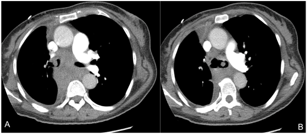
A 53-year-old woman presented to the emergency department with massive hemoptysis and respiratory failure. She had had history of intermittent hemoptysis and worsening shortness of breath during the last year. Her past medical history was remarkable for recently diagnosed lung cancer, two months previous to the admission. A metastatic poorly differentiated lung adenocarcinoma was reported in pathology from another medical center. On admission she required or tracheal intubation. The physical examination was, otherwise, unremarkable. She was hemodynamically stable and did not require vasopressor. Platelets and coagulation studies were in the normal range. A chest CT-scan with contrast showed evidence of a 6-cm-diameter right perihilar tumor, with extension to bilateral main-bronchi (Figure 1). There was secondary 70% left-main-bronchi obstruction and right-main-bronchi almost obliteration. Endobronchial obstruction due to advanced lung cancer was considered. A therapeutic bronchoscopy was performed. It showed 90% obstruction of bilateral main-bronchi by easily bleeding polypoid mass. Resection by cauterization was performed, allowing bleeding control and permeabilization of the airway. Samples were sent to pathology.

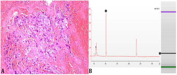
Bronchoalveolar lavage was negative for bacterial cultures, as well as for mycobacterial PCR, KOH, and Ziehl-Neelsen stain. The cytology reported inflammatory cells, with no evidence of malignant cells. The patient was extubated 24-hours after the bronchoscopy. Facing the absence of previous studies for lung cancer staging, complementary radiological imaging was done. The CT-brain with contrast showed no abnormality. A photopenic defect in the right sacroiliac-junction compatible with a metastatic lytic lesion was seen in a bone-scan. The abdominopelvic-MRI showed no significant findings. The pathology report of the resected tissue showed a malignant epithelial neoplasm composed of pleomorphic cells compatible with a pure choriocarcinoma (Figure 2A). Immunohistochemistry was positive for βhCG and negative for TTF-1 (anti-Thyroid Transcription Factor-1), p63, cytokeratin-20, and calretinin. A primary pulmonary or gastrointestinal neoplasm was ruled out. The tumor was diagnosed as a metastatic choriocarcinoma. The patient was G3P2A1. She had history of two male births, the last one 30 years ago. The miscarriage had been 20 years before the admission. Menopause had been at 45-year-old, with no abnormal vaginal bleeding afterwards. There was no history of gestational trophoblastic disease.

The plasma β-hCG level was 122.416 mIU/ml [0-2 mIU/ml]. The gynecological examination and pelvic MRI showed no evidence of disease.
In order to determine the gestational or nongestational tumor origin, PCR looking for Y chromosome specific regions was performed. The PCR amplified the SRY (sex-determining region Y), as well as the followings Short Tandem Repeats (STR): DYS19, DYS392, and DYS439. The molecular studies identified the presence of Y chromosome regions in the tumor. It confirmed a gestational type of metastatic choriocarcinoma (Figure 2B). She was started on chemotherapy. EMA/CO (etoposide, methotrexate, actinomycin D, cyclophosphamide, and vincristine) regimen was chosen. Clinical and radiological improvements were achieved. After three cycles of chemotherapy, the hCG went down to 93 mIU/ml. She received one more cycle of chemotherapy as an outpatient. On the follow-up, there was not further evidence of the disease with normal values of hCG.
Choriocarcinoma usually affects premenopausal women. A postmenopausal choriocarcinoma is a rare event and there are few cases reported in the literature. In our case, the tumor clinically was manifested 20years after the last pregnancy. The biological mechanism behind the long latent period for the gestational choriocarcinoma to develop in a postmenopausal woman has not been described. The gestational choriocarcinoma arises within the uterine cavity, however our case was presented with pulmonary metastatic involvement without evidence of uterine disease (i.e. spontaneous regression of the primary tumor). This behavior is known as the “burn-out” phenomenon. It has been described mainly in melanoma, as well as in other tumors including the germ cell tumors. A molecular immunological mechanism seems to be involved in this process. The tumor growth and spread cause repetitive exposures of the T-lymphocytes to the primary tumor antigens. The T-cells tend to eliminate the primary lesion, while the metastatic cells change the expression of the surface antigens. At the end, the activated immune response against the primary antigens is not effective for eliminating the metastatic cells, allowing them to stay.6 Despite of the lack of any uterine abnormality in the MRI, we cannot rule out the presence of microscopic neoplasm since the pathologic study was not done.
Gestational and non-gestational choriocarcinoma are clinically and pathologically indistinguishable. The only reliable technique to differentiate them is by genetic testing. In our report, the amplification of Y chromosome regions was sufficient to determine the gestational etiology. DNA polymorphism has been used to identify the genetic origin of choriocarcinoma with the analysis of STR mainly when the karyotype is XX.7
Determining the origin of choriocarcinoma is important to select the most appropriate treatment. The patient had stage IV choriocarcinoma according to FIGO, requiring an aggressive chemotherapeutic regimen. Low-risk and low-stage gestational choriocarcinoma could be treated with a single agent therapy (i.e. methotrexate). But, irrespective of the stage, nongestational choriocarcinoma should be treated with multi-agent chemotherapy due to its lower response to treatment and poor prognosis compared with the gestational type.8
We presented a metastatic gestational choriocarcinoma in a postmenopausal woman, manifested clinically 20 years after her last pregnancy. Systemic chemotherapy must be started immediately after the diagnosis, due to the high chance to achieve a complete response.
None.
The authors declare they have no conflict of interest. This study was carried out with resources from the Fundación Valle del Lili.

©2016 Fernandez, et al. This is an open access article distributed under the terms of the, which permits unrestricted use, distribution, and build upon your work non-commercially.