Advances in
eISSN: 2377-4290


Case Report Volume 10 Issue 2
1Consultant Vitreo-retina, Dhami eye care hospital, India
2Cornea and refractive consultant, Dhami eye care hospital, India
3Medical director, Dhami eye care hospital, India
Correspondence: Abhinav Dhami, Consultant Vitreo-retina, Dhami eye care hospital, 82-b kitchlu nagar, Ludhiana, Punjab, India, Tel +91-8220221044
Received: February 06, 2020 | Published: April 23, 2020
Citation: Dhami A, Dhami NB, Dhami GS. Spontaneous closure of stage 2 macular hole after cataract surgery. Adv Ophthalmol Vis Syst. 2020;10(2):45-46. DOI: 10.15406/aovs.2020.10.00381
We report a case of 64-year-old female, presented with decreased vision in left eye, for 3 months. Her vision was 20/30, N6 in right eye and 20/200 and N36 in left eye. She had nuclear sclerosis grade 2 and optical coherence tomography showed stage-2 macular hole in left eye. The macular hole diameter was 286 microns and the basal dimeter of 486 microns. The patient underwent cataract surgery with intraocular lens implantation in left eye. At 1 month, an inner retinal layer apposition was noted with subsequent inner to outer retinal apposition, resulting in complete spontaneous closure at the 3rd month follow up with visual acuity gain of 20/40, N8. This case highlights that stage-2 macular hole can close spontaneously with relief of vitreomacular traction.
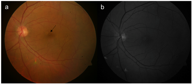
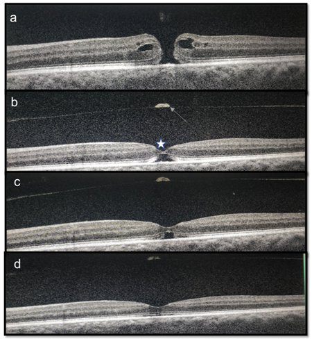
A 64-year-old female, presented with decreased vision in left eye, since 3 months. The visual acuity on Snellens chart was 20/30, N6 in right eye and 20/200 and N36 in left eye. Anterior segment examination showed both eyes with nuclear sclerosis of grade 3. The fundus of right eye was within normal limits with left eye optic disc and macula appeared normal (Figure1a &b). As vison was not correlating, optical coherence tomography (OCT) was done which revealed full thickness macular hole (FTMH) (Figure 2a). The dimensions of MH were, diameter of 286microns (m) and the basal dimeter of 486m. The patient was advised combined surgery but opted for only cataract extraction (phacoemulsification with a 2.8mm corneal incision) with single piece foldable intraocular lens implantation with no intraoperative complications. At 1 month follow up vision was 20/60, N24. OCT in left eye showed a separated vitreous with operculum with inner retinal layer apposition with outer lamellar hole (Figure 2b). At 2nd month, the inner retinal apposition increased, with decrease in outer lamella hole dimension (Figure 2c) and at 3rd month there was complete apposition of inner and outer retinal layers with final visual acuity of 20/40, N8 in left eye.

Figure 1 (a) Color fundus photograph of the left eye, black arrow points to greyish reflex of the separated posterior vitreous above the fovea. (b) red free photograph of the left eye.

Figure 2 (a) OCT showing a full thickness macular hole. (b) At 1 month post cataract surgery inner retinal layer apposition (white star) is noted with outer lamellar hole with posterior hyaloid separation(white line). (c) OCT at 2nd month, there is increase in inner retinal layer apposition with decrease in the size of outer lamellar hole. (d) OCT at 3rd month showing complete spontaneous apposition of the inner to outer retinal layers.
The spontaneous closure of idiopathic MH is less prevalent and less well understood with a handful of case reports in literature.1 Closure is also more likely when the hole is small(diameter size of less than 250μm)2 and in traumatic MH within first six months.3 Various mechanisms for the spontaneous closure of idiopathic MH described are a) complete detachment of the posterior hyaloid, leading to a reduction in antero-posterior tractional forces; b) bridging effect of retinal tissue and glial cell proliferation; c) formation of a contractile epiretinal membrane.4 The incidence of spontaneous idiopathic macular hole closure reported in literature is 2.7–6.2% and 11.5% of patients are associated with a posterior vitreous detachment. The minimum time to closure is three months to the definitive closure since the starting of the bridging effect as is reported in other similar report in the literature.1 Visual recovery is variable as it depends upon the period for which macular hole was present till its closure and the associated photoreceptor damage at the fovea, during the re-apposition of the inner and outer retinal layer.5 In our case, closure can be attributed due to posterior vitreous detachment after cataract surgery.6 The case highlights that small diameter MH can resolve without any retinal surgical intervention with a posterior vitreous separation.
None.
No financial funding disclosure.
No proprietary interest or conflict.

©2020 Dhami, et al. This is an open access article distributed under the terms of the, which permits unrestricted use, distribution, and build upon your work non-commercially.